{"title":"纳米级核组织体内研究的显微方法。","authors":"Nidhi Rani Lokesh, Mark E Pownall","doi":"10.1042/BST20240629","DOIUrl":null,"url":null,"abstract":"<p><p>Eukaryotic genomes are highly compacted within the nucleus and organized into complex 3D structures across various genomic and physical scales. Organization within the nucleus plays a key role in gene regulation, both facilitating regulatory interactions to promote transcription while also enabling the silencing of other genes. Despite the functional importance of genome organization in determining cell identity and function, investigating nuclear organization across this wide range of physical scales has been challenging. Microscopy provides the opportunity for direct visualization of nuclear structures and has pioneered key discoveries in this field. Nonetheless, visualization of nanoscale structures within the nucleus, such as nucleosomes and chromatin loops, requires super-resolution imaging to go beyond the ~220 nm diffraction limit. Here, we review recent advances in imaging technology and their promise to uncover new insights into the organization of the nucleus at the nanoscale. We discuss different imaging modalities and how they have been applied to the nucleus, with a focus on super-resolution light microscopy and its application to in vivo systems. Finally, we conclude with our perspective on how continued technical innovations in super-resolution imaging in the nucleus will advance our understanding of genome structure and function.</p>","PeriodicalId":8841,"journal":{"name":"Biochemical Society transactions","volume":"53 1","pages":""},"PeriodicalIF":4.3000,"publicationDate":"2025-02-03","publicationTypes":"Journal Article","fieldsOfStudy":null,"isOpenAccess":false,"openAccessPdf":"https://www.ncbi.nlm.nih.gov/pmc/articles/PMC12224908/pdf/","citationCount":"0","resultStr":"{\"title\":\"Microscopy methods for the in vivo study of nanoscale nuclear organization.\",\"authors\":\"Nidhi Rani Lokesh, Mark E Pownall\",\"doi\":\"10.1042/BST20240629\",\"DOIUrl\":null,\"url\":null,\"abstract\":\"<p><p>Eukaryotic genomes are highly compacted within the nucleus and organized into complex 3D structures across various genomic and physical scales. Organization within the nucleus plays a key role in gene regulation, both facilitating regulatory interactions to promote transcription while also enabling the silencing of other genes. Despite the functional importance of genome organization in determining cell identity and function, investigating nuclear organization across this wide range of physical scales has been challenging. Microscopy provides the opportunity for direct visualization of nuclear structures and has pioneered key discoveries in this field. Nonetheless, visualization of nanoscale structures within the nucleus, such as nucleosomes and chromatin loops, requires super-resolution imaging to go beyond the ~220 nm diffraction limit. Here, we review recent advances in imaging technology and their promise to uncover new insights into the organization of the nucleus at the nanoscale. We discuss different imaging modalities and how they have been applied to the nucleus, with a focus on super-resolution light microscopy and its application to in vivo systems. Finally, we conclude with our perspective on how continued technical innovations in super-resolution imaging in the nucleus will advance our understanding of genome structure and function.</p>\",\"PeriodicalId\":8841,\"journal\":{\"name\":\"Biochemical Society transactions\",\"volume\":\"53 1\",\"pages\":\"\"},\"PeriodicalIF\":4.3000,\"publicationDate\":\"2025-02-03\",\"publicationTypes\":\"Journal Article\",\"fieldsOfStudy\":null,\"isOpenAccess\":false,\"openAccessPdf\":\"https://www.ncbi.nlm.nih.gov/pmc/articles/PMC12224908/pdf/\",\"citationCount\":\"0\",\"resultStr\":null,\"platform\":\"Semanticscholar\",\"paperid\":null,\"PeriodicalName\":\"Biochemical Society transactions\",\"FirstCategoryId\":\"99\",\"ListUrlMain\":\"https://doi.org/10.1042/BST20240629\",\"RegionNum\":3,\"RegionCategory\":\"生物学\",\"ArticlePicture\":[],\"TitleCN\":null,\"AbstractTextCN\":null,\"PMCID\":null,\"EPubDate\":\"\",\"PubModel\":\"\",\"JCR\":\"Q2\",\"JCRName\":\"BIOCHEMISTRY & MOLECULAR BIOLOGY\",\"Score\":null,\"Total\":0}","platform":"Semanticscholar","paperid":null,"PeriodicalName":"Biochemical Society transactions","FirstCategoryId":"99","ListUrlMain":"https://doi.org/10.1042/BST20240629","RegionNum":3,"RegionCategory":"生物学","ArticlePicture":[],"TitleCN":null,"AbstractTextCN":null,"PMCID":null,"EPubDate":"","PubModel":"","JCR":"Q2","JCRName":"BIOCHEMISTRY & MOLECULAR BIOLOGY","Score":null,"Total":0}
Microscopy methods for the in vivo study of nanoscale nuclear organization.
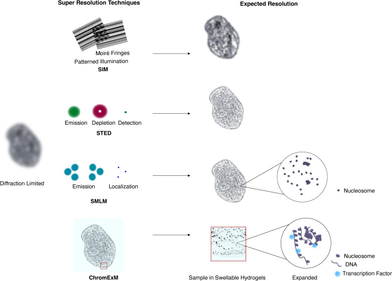
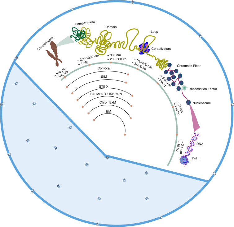
Eukaryotic genomes are highly compacted within the nucleus and organized into complex 3D structures across various genomic and physical scales. Organization within the nucleus plays a key role in gene regulation, both facilitating regulatory interactions to promote transcription while also enabling the silencing of other genes. Despite the functional importance of genome organization in determining cell identity and function, investigating nuclear organization across this wide range of physical scales has been challenging. Microscopy provides the opportunity for direct visualization of nuclear structures and has pioneered key discoveries in this field. Nonetheless, visualization of nanoscale structures within the nucleus, such as nucleosomes and chromatin loops, requires super-resolution imaging to go beyond the ~220 nm diffraction limit. Here, we review recent advances in imaging technology and their promise to uncover new insights into the organization of the nucleus at the nanoscale. We discuss different imaging modalities and how they have been applied to the nucleus, with a focus on super-resolution light microscopy and its application to in vivo systems. Finally, we conclude with our perspective on how continued technical innovations in super-resolution imaging in the nucleus will advance our understanding of genome structure and function.
期刊介绍:
Biochemical Society Transactions is the reviews journal of the Biochemical Society. Publishing concise reviews written by experts in the field, providing a timely snapshot of the latest developments across all areas of the molecular and cellular biosciences.
Elevating our authors’ ideas and expertise, each review includes a perspectives section where authors offer comment on the latest advances, a glimpse of future challenges and highlighting the importance of associated research areas in far broader contexts.
 求助内容:
求助内容: 应助结果提醒方式:


