Martin Tripon, Matthieu Lalevee, Floris van Rooij, Chinyelum Agu, Mo Saffarini, Philippe Beaudet
{"title":"拇外翻足前、中足角的3DCT标准负重和籽骨位比较。","authors":"Martin Tripon, Matthieu Lalevee, Floris van Rooij, Chinyelum Agu, Mo Saffarini, Philippe Beaudet","doi":"10.1302/2046-3758.142.BJR-2024-0172.R2","DOIUrl":null,"url":null,"abstract":"<p><strong>Aims: </strong>To evaluate how fore- and midfoot coronal plane alignment differs in feet with hallux valgus (HV), using 3DCT when measured in standard weightbearing (SWB) versus sesamoid view (SV) position, and to determine whether first metatarsophalangeal (MTP) dorsiflexion affects the relationship between the first metatarsal (M1) head and the sesamoid bones.</p><p><strong>Methods: </strong>A consecutive series of 34 feet that underwent 3DCT in SWB and SV positions for symptomatic HV was assessed, of which four feet were excluded for distorted or incomplete images. Two foot and ankle clinicians independently digitized a series of points, and measured a series of angles according to a pre-defined protocol. Measurements include navicular pronation angle, M1 head (Saltzman angle), and metatarsosesamoid rotation angle (MSRA).</p><p><strong>Results: </strong>The mean age of the 30 patients was 57.5 years (SD 13.4). The mean navicular pronation angle was significantly smaller in the SV position (9.6° (SD 4.4°)) compared to the SWB position (16.4° (SD 5.8°); p < 0.001). There was a difference in MSRA between the SWB and SV positions, revealing an increase in MSRA in 22 patients, while there was a decrease in eight patients. In patients where the MSRA increased, the mean Saltzman angle was 2.5° (SD 5.7°) lower in the SV position versus the SWB position, while in patients where MSRA decreased, the mean Saltzman angle was 3.4° (SD 3.6°) greater in the SV position versus the SWB position.</p><p><strong>Conclusion: </strong>MTP dorsiflexion causes supination of the navicular, while other first ray parameters remain unchanged, and has a greater influence on the M1 head coronal alignment than on the sesamoids. MTP dorsiflexion induces axial rotations of M1, which vary in direction and magnitude from one patient to another.</p>","PeriodicalId":9074,"journal":{"name":"Bone & Joint Research","volume":"14 2","pages":"69-76"},"PeriodicalIF":5.1000,"publicationDate":"2025-02-01","publicationTypes":"Journal Article","fieldsOfStudy":null,"isOpenAccess":false,"openAccessPdf":"https://www.ncbi.nlm.nih.gov/pmc/articles/PMC11785418/pdf/","citationCount":"0","resultStr":"{\"title\":\"Comparison of fore- and midfoot angles using 3DCT in standard weightbearing and sesamoid view position in feet with hallux valgus.\",\"authors\":\"Martin Tripon, Matthieu Lalevee, Floris van Rooij, Chinyelum Agu, Mo Saffarini, Philippe Beaudet\",\"doi\":\"10.1302/2046-3758.142.BJR-2024-0172.R2\",\"DOIUrl\":null,\"url\":null,\"abstract\":\"<p><strong>Aims: </strong>To evaluate how fore- and midfoot coronal plane alignment differs in feet with hallux valgus (HV), using 3DCT when measured in standard weightbearing (SWB) versus sesamoid view (SV) position, and to determine whether first metatarsophalangeal (MTP) dorsiflexion affects the relationship between the first metatarsal (M1) head and the sesamoid bones.</p><p><strong>Methods: </strong>A consecutive series of 34 feet that underwent 3DCT in SWB and SV positions for symptomatic HV was assessed, of which four feet were excluded for distorted or incomplete images. Two foot and ankle clinicians independently digitized a series of points, and measured a series of angles according to a pre-defined protocol. Measurements include navicular pronation angle, M1 head (Saltzman angle), and metatarsosesamoid rotation angle (MSRA).</p><p><strong>Results: </strong>The mean age of the 30 patients was 57.5 years (SD 13.4). The mean navicular pronation angle was significantly smaller in the SV position (9.6° (SD 4.4°)) compared to the SWB position (16.4° (SD 5.8°); p < 0.001). There was a difference in MSRA between the SWB and SV positions, revealing an increase in MSRA in 22 patients, while there was a decrease in eight patients. In patients where the MSRA increased, the mean Saltzman angle was 2.5° (SD 5.7°) lower in the SV position versus the SWB position, while in patients where MSRA decreased, the mean Saltzman angle was 3.4° (SD 3.6°) greater in the SV position versus the SWB position.</p><p><strong>Conclusion: </strong>MTP dorsiflexion causes supination of the navicular, while other first ray parameters remain unchanged, and has a greater influence on the M1 head coronal alignment than on the sesamoids. MTP dorsiflexion induces axial rotations of M1, which vary in direction and magnitude from one patient to another.</p>\",\"PeriodicalId\":9074,\"journal\":{\"name\":\"Bone & Joint Research\",\"volume\":\"14 2\",\"pages\":\"69-76\"},\"PeriodicalIF\":5.1000,\"publicationDate\":\"2025-02-01\",\"publicationTypes\":\"Journal Article\",\"fieldsOfStudy\":null,\"isOpenAccess\":false,\"openAccessPdf\":\"https://www.ncbi.nlm.nih.gov/pmc/articles/PMC11785418/pdf/\",\"citationCount\":\"0\",\"resultStr\":null,\"platform\":\"Semanticscholar\",\"paperid\":null,\"PeriodicalName\":\"Bone & Joint Research\",\"FirstCategoryId\":\"3\",\"ListUrlMain\":\"https://doi.org/10.1302/2046-3758.142.BJR-2024-0172.R2\",\"RegionNum\":2,\"RegionCategory\":\"医学\",\"ArticlePicture\":[],\"TitleCN\":null,\"AbstractTextCN\":null,\"PMCID\":null,\"EPubDate\":\"\",\"PubModel\":\"\",\"JCR\":\"Q2\",\"JCRName\":\"CELL & TISSUE ENGINEERING\",\"Score\":null,\"Total\":0}","platform":"Semanticscholar","paperid":null,"PeriodicalName":"Bone & Joint Research","FirstCategoryId":"3","ListUrlMain":"https://doi.org/10.1302/2046-3758.142.BJR-2024-0172.R2","RegionNum":2,"RegionCategory":"医学","ArticlePicture":[],"TitleCN":null,"AbstractTextCN":null,"PMCID":null,"EPubDate":"","PubModel":"","JCR":"Q2","JCRName":"CELL & TISSUE ENGINEERING","Score":null,"Total":0}
Comparison of fore- and midfoot angles using 3DCT in standard weightbearing and sesamoid view position in feet with hallux valgus.
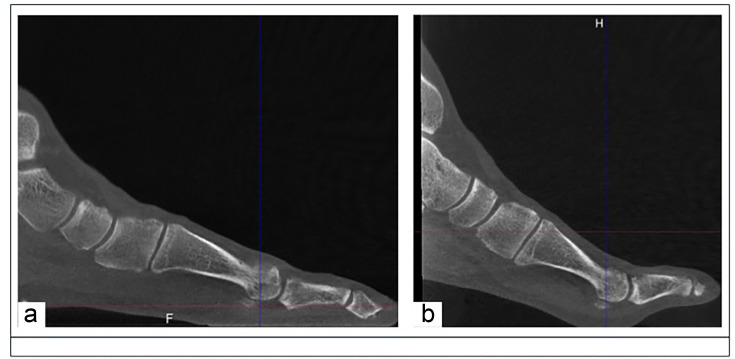
Aims: To evaluate how fore- and midfoot coronal plane alignment differs in feet with hallux valgus (HV), using 3DCT when measured in standard weightbearing (SWB) versus sesamoid view (SV) position, and to determine whether first metatarsophalangeal (MTP) dorsiflexion affects the relationship between the first metatarsal (M1) head and the sesamoid bones.
Methods: A consecutive series of 34 feet that underwent 3DCT in SWB and SV positions for symptomatic HV was assessed, of which four feet were excluded for distorted or incomplete images. Two foot and ankle clinicians independently digitized a series of points, and measured a series of angles according to a pre-defined protocol. Measurements include navicular pronation angle, M1 head (Saltzman angle), and metatarsosesamoid rotation angle (MSRA).
Results: The mean age of the 30 patients was 57.5 years (SD 13.4). The mean navicular pronation angle was significantly smaller in the SV position (9.6° (SD 4.4°)) compared to the SWB position (16.4° (SD 5.8°); p < 0.001). There was a difference in MSRA between the SWB and SV positions, revealing an increase in MSRA in 22 patients, while there was a decrease in eight patients. In patients where the MSRA increased, the mean Saltzman angle was 2.5° (SD 5.7°) lower in the SV position versus the SWB position, while in patients where MSRA decreased, the mean Saltzman angle was 3.4° (SD 3.6°) greater in the SV position versus the SWB position.
Conclusion: MTP dorsiflexion causes supination of the navicular, while other first ray parameters remain unchanged, and has a greater influence on the M1 head coronal alignment than on the sesamoids. MTP dorsiflexion induces axial rotations of M1, which vary in direction and magnitude from one patient to another.
 求助内容:
求助内容: 应助结果提醒方式:


