Kimberly A Nickerson, Christina Carranza, Scott Telfer, William R Ledoux, Brittney C Muir
{"title":"三维差异的足底表面形状捕获的方法用于定制适应性鞋垫设计。","authors":"Kimberly A Nickerson, Christina Carranza, Scott Telfer, William R Ledoux, Brittney C Muir","doi":"10.1002/jfa2.70034","DOIUrl":null,"url":null,"abstract":"<p><strong>Background: </strong>The patient-specific shape of custom accommodative insoles for individuals with diabetes provides full foot-to-insole contact, offloading areas with high plantar pressures and reducing ulceration risk. To design the insole surface, plantar surface shape is captured, traditionally with a foam crush box impression or more recently with 3D scans of the foot. Beyond discrete measurements of the foot, the overall plantar surface shapes obtained from these different methods have yet to be compared, however, differences in the shapes captured by these methods may affect the insole's surface geometry design and subsequent performance.</p><p><strong>Methods: </strong>Plantar surface shapes of 12 individuals with diabetes were captured using a foam crush box, flatbed 3D foot scanner, and handheld 3D scanner. Foot length, width, arch height, and arch volume were measured from each shape-capture method and compared. Mesh-to-mesh distances between the foam crush box mesh and the direct scanning method meshes for each subject were calculated.</p><p><strong>Results: </strong>Foot length and width measured from the foam crush box scan were greater than the foot length measured from the flatbed scan and handheld scan. The flatbed scan also measured a length and width greater than the handheld scan. Arch heights and volumes from the flatbed scan were less than the heights calculated from the foam crush box and handheld scan. Mesh-to-mesh distances for the flatbed scan and areas of the foot not in contact with the scanner were inferior to the corresponding areas in the foam crush box impression. For the handheld scan, the lateral hindfoot and midfoot were superior, and the medial forefoot was inferior to the foam crush box impression.</p><p><strong>Conclusions: </strong>Different clinical methods used to capture foot shapes for the design of accommodative insoles may result in different plantar surface shape outputs and therefore impact custom accommodative insole design.</p>","PeriodicalId":49164,"journal":{"name":"Journal of Foot and Ankle Research","volume":"18 1","pages":"e70034"},"PeriodicalIF":2.2000,"publicationDate":"2025-03-01","publicationTypes":"Journal Article","fieldsOfStudy":null,"isOpenAccess":false,"openAccessPdf":"https://www.ncbi.nlm.nih.gov/pmc/articles/PMC11781946/pdf/","citationCount":"0","resultStr":"{\"title\":\"Three-dimensional differences in plantar surface shape captured by methods used for custom accommodative insole design.\",\"authors\":\"Kimberly A Nickerson, Christina Carranza, Scott Telfer, William R Ledoux, Brittney C Muir\",\"doi\":\"10.1002/jfa2.70034\",\"DOIUrl\":null,\"url\":null,\"abstract\":\"<p><strong>Background: </strong>The patient-specific shape of custom accommodative insoles for individuals with diabetes provides full foot-to-insole contact, offloading areas with high plantar pressures and reducing ulceration risk. To design the insole surface, plantar surface shape is captured, traditionally with a foam crush box impression or more recently with 3D scans of the foot. Beyond discrete measurements of the foot, the overall plantar surface shapes obtained from these different methods have yet to be compared, however, differences in the shapes captured by these methods may affect the insole's surface geometry design and subsequent performance.</p><p><strong>Methods: </strong>Plantar surface shapes of 12 individuals with diabetes were captured using a foam crush box, flatbed 3D foot scanner, and handheld 3D scanner. Foot length, width, arch height, and arch volume were measured from each shape-capture method and compared. Mesh-to-mesh distances between the foam crush box mesh and the direct scanning method meshes for each subject were calculated.</p><p><strong>Results: </strong>Foot length and width measured from the foam crush box scan were greater than the foot length measured from the flatbed scan and handheld scan. The flatbed scan also measured a length and width greater than the handheld scan. Arch heights and volumes from the flatbed scan were less than the heights calculated from the foam crush box and handheld scan. Mesh-to-mesh distances for the flatbed scan and areas of the foot not in contact with the scanner were inferior to the corresponding areas in the foam crush box impression. For the handheld scan, the lateral hindfoot and midfoot were superior, and the medial forefoot was inferior to the foam crush box impression.</p><p><strong>Conclusions: </strong>Different clinical methods used to capture foot shapes for the design of accommodative insoles may result in different plantar surface shape outputs and therefore impact custom accommodative insole design.</p>\",\"PeriodicalId\":49164,\"journal\":{\"name\":\"Journal of Foot and Ankle Research\",\"volume\":\"18 1\",\"pages\":\"e70034\"},\"PeriodicalIF\":2.2000,\"publicationDate\":\"2025-03-01\",\"publicationTypes\":\"Journal Article\",\"fieldsOfStudy\":null,\"isOpenAccess\":false,\"openAccessPdf\":\"https://www.ncbi.nlm.nih.gov/pmc/articles/PMC11781946/pdf/\",\"citationCount\":\"0\",\"resultStr\":null,\"platform\":\"Semanticscholar\",\"paperid\":null,\"PeriodicalName\":\"Journal of Foot and Ankle Research\",\"FirstCategoryId\":\"3\",\"ListUrlMain\":\"https://doi.org/10.1002/jfa2.70034\",\"RegionNum\":3,\"RegionCategory\":\"医学\",\"ArticlePicture\":[],\"TitleCN\":null,\"AbstractTextCN\":null,\"PMCID\":null,\"EPubDate\":\"\",\"PubModel\":\"\",\"JCR\":\"Q1\",\"JCRName\":\"ORTHOPEDICS\",\"Score\":null,\"Total\":0}","platform":"Semanticscholar","paperid":null,"PeriodicalName":"Journal of Foot and Ankle Research","FirstCategoryId":"3","ListUrlMain":"https://doi.org/10.1002/jfa2.70034","RegionNum":3,"RegionCategory":"医学","ArticlePicture":[],"TitleCN":null,"AbstractTextCN":null,"PMCID":null,"EPubDate":"","PubModel":"","JCR":"Q1","JCRName":"ORTHOPEDICS","Score":null,"Total":0}
Three-dimensional differences in plantar surface shape captured by methods used for custom accommodative insole design.
Background: The patient-specific shape of custom accommodative insoles for individuals with diabetes provides full foot-to-insole contact, offloading areas with high plantar pressures and reducing ulceration risk. To design the insole surface, plantar surface shape is captured, traditionally with a foam crush box impression or more recently with 3D scans of the foot. Beyond discrete measurements of the foot, the overall plantar surface shapes obtained from these different methods have yet to be compared, however, differences in the shapes captured by these methods may affect the insole's surface geometry design and subsequent performance.
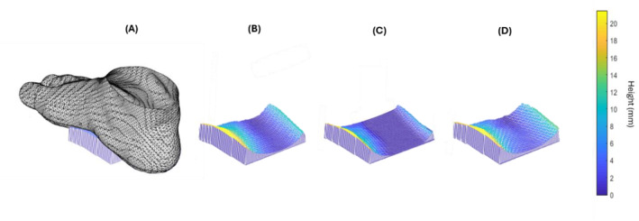
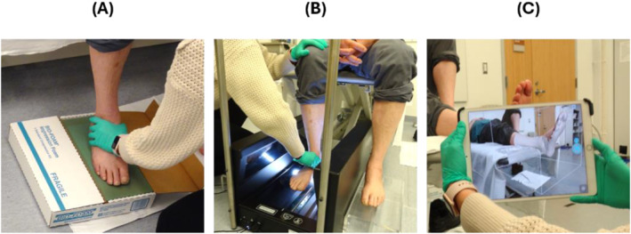
Methods: Plantar surface shapes of 12 individuals with diabetes were captured using a foam crush box, flatbed 3D foot scanner, and handheld 3D scanner. Foot length, width, arch height, and arch volume were measured from each shape-capture method and compared. Mesh-to-mesh distances between the foam crush box mesh and the direct scanning method meshes for each subject were calculated.
Results: Foot length and width measured from the foam crush box scan were greater than the foot length measured from the flatbed scan and handheld scan. The flatbed scan also measured a length and width greater than the handheld scan. Arch heights and volumes from the flatbed scan were less than the heights calculated from the foam crush box and handheld scan. Mesh-to-mesh distances for the flatbed scan and areas of the foot not in contact with the scanner were inferior to the corresponding areas in the foam crush box impression. For the handheld scan, the lateral hindfoot and midfoot were superior, and the medial forefoot was inferior to the foam crush box impression.
Conclusions: Different clinical methods used to capture foot shapes for the design of accommodative insoles may result in different plantar surface shape outputs and therefore impact custom accommodative insole design.
期刊介绍:
Journal of Foot and Ankle Research, the official journal of the Australian Podiatry Association and The College of Podiatry (UK), is an open access journal that encompasses all aspects of policy, organisation, delivery and clinical practice related to the assessment, diagnosis, prevention and management of foot and ankle disorders.
Journal of Foot and Ankle Research covers a wide range of clinical subject areas, including diabetology, paediatrics, sports medicine, gerontology and geriatrics, foot surgery, physical therapy, dermatology, wound management, radiology, biomechanics and bioengineering, orthotics and prosthetics, as well the broad areas of epidemiology, policy, organisation and delivery of services related to foot and ankle care.
The journal encourages submissions from all health professionals who manage lower limb conditions, including podiatrists, nurses, physical therapists and physiotherapists, orthopaedists, manual therapists, medical specialists and general medical practitioners, as well as health service researchers concerned with foot and ankle care.
The Australian Podiatry Association and the College of Podiatry (UK) have reserve funds to cover the article-processing charge for manuscripts submitted by its members. Society members can email the appropriate contact at Australian Podiatry Association or The College of Podiatry to obtain the corresponding code to enter on submission.
 求助内容:
求助内容: 应助结果提醒方式:


