Andressa Colares da Costa Otavio, Mariana Vicereki Trescastro, Hilton Justino da Silva, Erissandra Gomes, Têmis Maria Félix
{"title":"儿童和青少年成骨不全症的咬肌和颞前肌的咀嚼和电激活。","authors":"Andressa Colares da Costa Otavio, Mariana Vicereki Trescastro, Hilton Justino da Silva, Erissandra Gomes, Têmis Maria Félix","doi":"10.1590/2317-1782/e20240052en","DOIUrl":null,"url":null,"abstract":"<p><strong>Purpose: </strong>to characterize mastication and electrical activation of the masseter and anterior temporalis muscles in children and adolescents with osteogenesis imperfecta (OI), and relate results to guided occlusion and occlusal interference.</p><p><strong>Methods: </strong>This observational, analytical cross-sectional study included 22 subjects divided into mild OI (MOI) (type 1) (n=15) and moderate-to-severe OI (MSOI) (types 3, 4, and 5) (n=7) groups. The Orofacial Myofunctional Evaluation with Scores (OMES) form was used to evaluate the clinical aspects of mastication. Surface electromyography was performed on the masseter and anterior temporalis muscles at rest for 10 seconds and during maximum intercuspation, spontaneous chewing, and instructed chewing on the right and left sides. Additionally, the activation index and muscle symmetry were measured.</p><p><strong>Results: </strong>a preferentially unilateral chewing pattern was observed in 12 (54.5%) participants. Masticatory patterns did not influence electrical activation during any of the tasks, nor did occlusal guidance during maximum intercuspation or mastication. The percentage of muscle activation during maximal intercuspation approached half of the total activation during spontaneous chewing. In muscle activation indices, the MSOI group presented more atypical scores, while the MOI group scores seemed to be in line with reference values. The symmetry indices seemed to correspond to reference values, but the standard deviation and minimum and maximum values pointed to asymmetric results.</p><p><strong>Conclusion: </strong>This study found that the OI population presented muscle imbalances, but the results did not allow us to define one pattern of change.</p>","PeriodicalId":46547,"journal":{"name":"CoDAS","volume":"37 1","pages":"e20240052"},"PeriodicalIF":0.8000,"publicationDate":"2025-01-27","publicationTypes":"Journal Article","fieldsOfStudy":null,"isOpenAccess":false,"openAccessPdf":"https://www.ncbi.nlm.nih.gov/pmc/articles/PMC11781364/pdf/","citationCount":"0","resultStr":"{\"title\":\"Mastication and electrical activation in the masseter and anterior temporalis muscles of children and adolescents with osteogenesis imperfecta.\",\"authors\":\"Andressa Colares da Costa Otavio, Mariana Vicereki Trescastro, Hilton Justino da Silva, Erissandra Gomes, Têmis Maria Félix\",\"doi\":\"10.1590/2317-1782/e20240052en\",\"DOIUrl\":null,\"url\":null,\"abstract\":\"<p><strong>Purpose: </strong>to characterize mastication and electrical activation of the masseter and anterior temporalis muscles in children and adolescents with osteogenesis imperfecta (OI), and relate results to guided occlusion and occlusal interference.</p><p><strong>Methods: </strong>This observational, analytical cross-sectional study included 22 subjects divided into mild OI (MOI) (type 1) (n=15) and moderate-to-severe OI (MSOI) (types 3, 4, and 5) (n=7) groups. The Orofacial Myofunctional Evaluation with Scores (OMES) form was used to evaluate the clinical aspects of mastication. Surface electromyography was performed on the masseter and anterior temporalis muscles at rest for 10 seconds and during maximum intercuspation, spontaneous chewing, and instructed chewing on the right and left sides. Additionally, the activation index and muscle symmetry were measured.</p><p><strong>Results: </strong>a preferentially unilateral chewing pattern was observed in 12 (54.5%) participants. Masticatory patterns did not influence electrical activation during any of the tasks, nor did occlusal guidance during maximum intercuspation or mastication. The percentage of muscle activation during maximal intercuspation approached half of the total activation during spontaneous chewing. In muscle activation indices, the MSOI group presented more atypical scores, while the MOI group scores seemed to be in line with reference values. The symmetry indices seemed to correspond to reference values, but the standard deviation and minimum and maximum values pointed to asymmetric results.</p><p><strong>Conclusion: </strong>This study found that the OI population presented muscle imbalances, but the results did not allow us to define one pattern of change.</p>\",\"PeriodicalId\":46547,\"journal\":{\"name\":\"CoDAS\",\"volume\":\"37 1\",\"pages\":\"e20240052\"},\"PeriodicalIF\":0.8000,\"publicationDate\":\"2025-01-27\",\"publicationTypes\":\"Journal Article\",\"fieldsOfStudy\":null,\"isOpenAccess\":false,\"openAccessPdf\":\"https://www.ncbi.nlm.nih.gov/pmc/articles/PMC11781364/pdf/\",\"citationCount\":\"0\",\"resultStr\":null,\"platform\":\"Semanticscholar\",\"paperid\":null,\"PeriodicalName\":\"CoDAS\",\"FirstCategoryId\":\"1085\",\"ListUrlMain\":\"https://doi.org/10.1590/2317-1782/e20240052en\",\"RegionNum\":0,\"RegionCategory\":null,\"ArticlePicture\":[],\"TitleCN\":null,\"AbstractTextCN\":null,\"PMCID\":null,\"EPubDate\":\"2025/1/1 0:00:00\",\"PubModel\":\"eCollection\",\"JCR\":\"Q4\",\"JCRName\":\"AUDIOLOGY & SPEECH-LANGUAGE PATHOLOGY\",\"Score\":null,\"Total\":0}","platform":"Semanticscholar","paperid":null,"PeriodicalName":"CoDAS","FirstCategoryId":"1085","ListUrlMain":"https://doi.org/10.1590/2317-1782/e20240052en","RegionNum":0,"RegionCategory":null,"ArticlePicture":[],"TitleCN":null,"AbstractTextCN":null,"PMCID":null,"EPubDate":"2025/1/1 0:00:00","PubModel":"eCollection","JCR":"Q4","JCRName":"AUDIOLOGY & SPEECH-LANGUAGE PATHOLOGY","Score":null,"Total":0}
Mastication and electrical activation in the masseter and anterior temporalis muscles of children and adolescents with osteogenesis imperfecta.
Purpose: to characterize mastication and electrical activation of the masseter and anterior temporalis muscles in children and adolescents with osteogenesis imperfecta (OI), and relate results to guided occlusion and occlusal interference.
Methods: This observational, analytical cross-sectional study included 22 subjects divided into mild OI (MOI) (type 1) (n=15) and moderate-to-severe OI (MSOI) (types 3, 4, and 5) (n=7) groups. The Orofacial Myofunctional Evaluation with Scores (OMES) form was used to evaluate the clinical aspects of mastication. Surface electromyography was performed on the masseter and anterior temporalis muscles at rest for 10 seconds and during maximum intercuspation, spontaneous chewing, and instructed chewing on the right and left sides. Additionally, the activation index and muscle symmetry were measured.
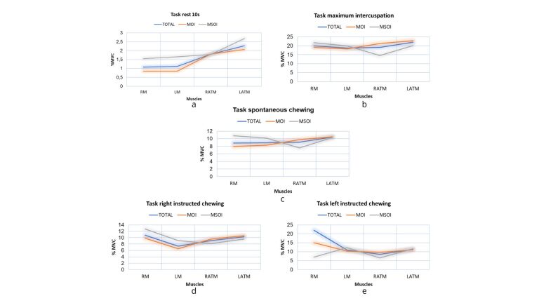
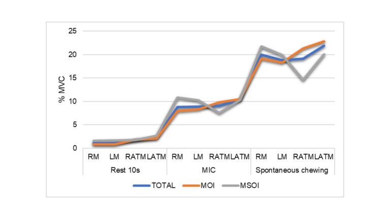
Results: a preferentially unilateral chewing pattern was observed in 12 (54.5%) participants. Masticatory patterns did not influence electrical activation during any of the tasks, nor did occlusal guidance during maximum intercuspation or mastication. The percentage of muscle activation during maximal intercuspation approached half of the total activation during spontaneous chewing. In muscle activation indices, the MSOI group presented more atypical scores, while the MOI group scores seemed to be in line with reference values. The symmetry indices seemed to correspond to reference values, but the standard deviation and minimum and maximum values pointed to asymmetric results.
Conclusion: This study found that the OI population presented muscle imbalances, but the results did not allow us to define one pattern of change.

 求助内容:
求助内容: 应助结果提醒方式:


