Jia-Qi Chen, Min Zheng, Wei Li, Jiong Zhou, Xiao-Yong Man
{"title":"硬皮病表皮角质形成细胞中天花素及其信号通路的调控。","authors":"Jia-Qi Chen, Min Zheng, Wei Li, Jiong Zhou, Xiao-Yong Man","doi":"10.5114/ada.2024.144841","DOIUrl":null,"url":null,"abstract":"<p><strong>Introduction: </strong>Systemic sclerosis is a complex disease characterized by the fibrosis and vasculopathy.</p><p><strong>Aim: </strong>We aimed to assess scleroderma by examining involucrin, an early terminal differentiation marker of epidermal keratinocytes.</p><p><strong>Material and methods: </strong>Immunolocalization of involucrin was performed in healthy controls and patients with scleroderma lesions by using an immunofluorescence (IF) assay. Normal and scleroderma keratinocytes were incubated in low-Ca basal-defined K-SFM overnight. Cells were pre-treated with Y-27632 (a Rho signalling pathway inhibitor) or interleukins (ILs), tumor necrosis factor-α (TNF-α), transforming growth factor β1 (TGF-β1), endothelin-1 (ET-1), interferon-γ (IFN)-γ, vascular endothelial growth factor (VEGF) and platelet-derived growth factor (PDGF) - BB. The involucrin protein expression level was measured by western blot.</p><p><strong>Results: </strong>Compared with the normal skin, involucrin was detected in the granular layer and the upper stratum spinosum in patients with scleroderma lesions. However, involucrin protein expression was decreased in cultured scleroderma keratinocytes. IL-6, IL-10, IL-17A, TNF-α, TGF-β1, ET-1, IFN-γ, VEGF165, and PDGF-BB increased involucrin expression in normal keratinocytes but decreased it in scleroderma keratinocytes. IFN-γ and IL-13 remarkably downregulated involucrin levels in normal and scleroderma keratinocytes, respectively, by Y-27632 intervention.</p><p><strong>Conclusions: </strong>Inflammatory cytokines, such as IL-6, IL-10, TGF-β1, ET-1, IFN-γ, VEGF, PDGF-BB, and Rho signalling pathway may be associated with the delayed epidermal differentiation in scleroderma.</p>","PeriodicalId":54595,"journal":{"name":"Postepy Dermatologii I Alergologii","volume":"41 6","pages":"604-609"},"PeriodicalIF":1.4000,"publicationDate":"2024-12-01","publicationTypes":"Journal Article","fieldsOfStudy":null,"isOpenAccess":false,"openAccessPdf":"https://www.ncbi.nlm.nih.gov/pmc/articles/PMC11770575/pdf/","citationCount":"0","resultStr":"{\"title\":\"Regulation of involucrin and signalling pathway in scleroderma epidermal keratinocytes.\",\"authors\":\"Jia-Qi Chen, Min Zheng, Wei Li, Jiong Zhou, Xiao-Yong Man\",\"doi\":\"10.5114/ada.2024.144841\",\"DOIUrl\":null,\"url\":null,\"abstract\":\"<p><strong>Introduction: </strong>Systemic sclerosis is a complex disease characterized by the fibrosis and vasculopathy.</p><p><strong>Aim: </strong>We aimed to assess scleroderma by examining involucrin, an early terminal differentiation marker of epidermal keratinocytes.</p><p><strong>Material and methods: </strong>Immunolocalization of involucrin was performed in healthy controls and patients with scleroderma lesions by using an immunofluorescence (IF) assay. Normal and scleroderma keratinocytes were incubated in low-Ca basal-defined K-SFM overnight. Cells were pre-treated with Y-27632 (a Rho signalling pathway inhibitor) or interleukins (ILs), tumor necrosis factor-α (TNF-α), transforming growth factor β1 (TGF-β1), endothelin-1 (ET-1), interferon-γ (IFN)-γ, vascular endothelial growth factor (VEGF) and platelet-derived growth factor (PDGF) - BB. The involucrin protein expression level was measured by western blot.</p><p><strong>Results: </strong>Compared with the normal skin, involucrin was detected in the granular layer and the upper stratum spinosum in patients with scleroderma lesions. However, involucrin protein expression was decreased in cultured scleroderma keratinocytes. IL-6, IL-10, IL-17A, TNF-α, TGF-β1, ET-1, IFN-γ, VEGF165, and PDGF-BB increased involucrin expression in normal keratinocytes but decreased it in scleroderma keratinocytes. IFN-γ and IL-13 remarkably downregulated involucrin levels in normal and scleroderma keratinocytes, respectively, by Y-27632 intervention.</p><p><strong>Conclusions: </strong>Inflammatory cytokines, such as IL-6, IL-10, TGF-β1, ET-1, IFN-γ, VEGF, PDGF-BB, and Rho signalling pathway may be associated with the delayed epidermal differentiation in scleroderma.</p>\",\"PeriodicalId\":54595,\"journal\":{\"name\":\"Postepy Dermatologii I Alergologii\",\"volume\":\"41 6\",\"pages\":\"604-609\"},\"PeriodicalIF\":1.4000,\"publicationDate\":\"2024-12-01\",\"publicationTypes\":\"Journal Article\",\"fieldsOfStudy\":null,\"isOpenAccess\":false,\"openAccessPdf\":\"https://www.ncbi.nlm.nih.gov/pmc/articles/PMC11770575/pdf/\",\"citationCount\":\"0\",\"resultStr\":null,\"platform\":\"Semanticscholar\",\"paperid\":null,\"PeriodicalName\":\"Postepy Dermatologii I Alergologii\",\"FirstCategoryId\":\"3\",\"ListUrlMain\":\"https://doi.org/10.5114/ada.2024.144841\",\"RegionNum\":4,\"RegionCategory\":\"医学\",\"ArticlePicture\":[],\"TitleCN\":null,\"AbstractTextCN\":null,\"PMCID\":null,\"EPubDate\":\"2024/11/8 0:00:00\",\"PubModel\":\"Epub\",\"JCR\":\"Q3\",\"JCRName\":\"ALLERGY\",\"Score\":null,\"Total\":0}","platform":"Semanticscholar","paperid":null,"PeriodicalName":"Postepy Dermatologii I Alergologii","FirstCategoryId":"3","ListUrlMain":"https://doi.org/10.5114/ada.2024.144841","RegionNum":4,"RegionCategory":"医学","ArticlePicture":[],"TitleCN":null,"AbstractTextCN":null,"PMCID":null,"EPubDate":"2024/11/8 0:00:00","PubModel":"Epub","JCR":"Q3","JCRName":"ALLERGY","Score":null,"Total":0}
Regulation of involucrin and signalling pathway in scleroderma epidermal keratinocytes.
Introduction: Systemic sclerosis is a complex disease characterized by the fibrosis and vasculopathy.
Aim: We aimed to assess scleroderma by examining involucrin, an early terminal differentiation marker of epidermal keratinocytes.
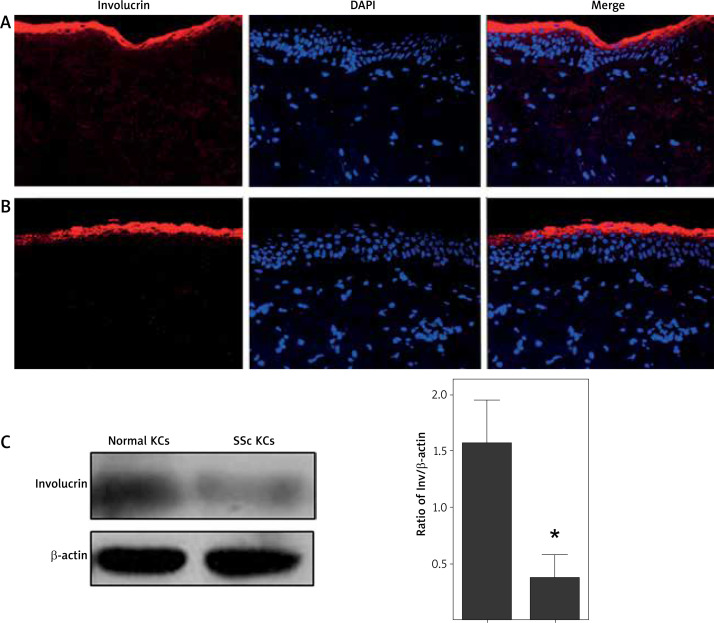
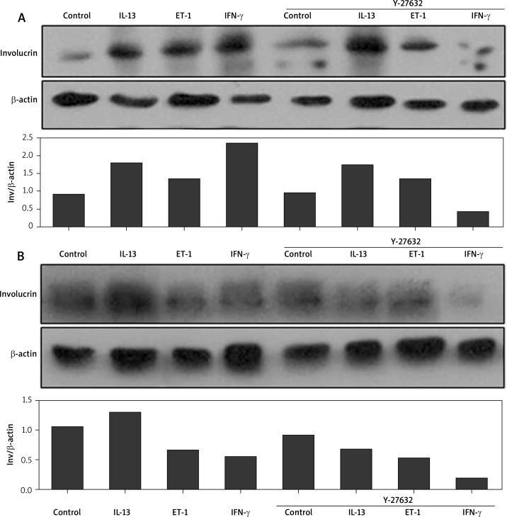
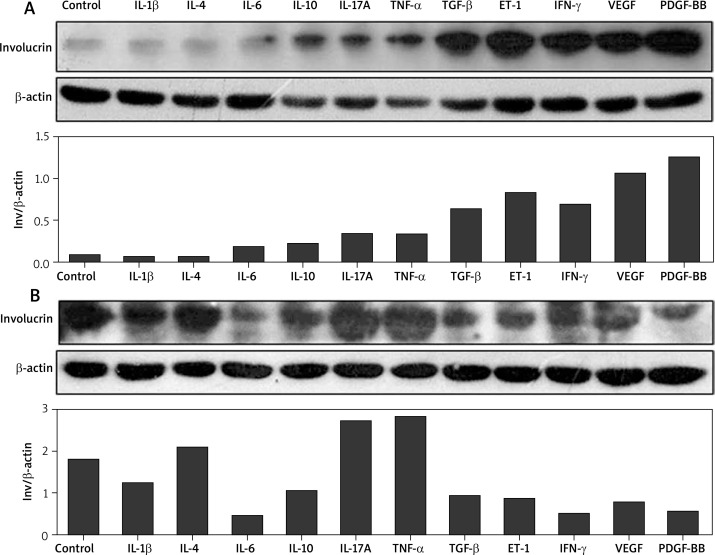
Material and methods: Immunolocalization of involucrin was performed in healthy controls and patients with scleroderma lesions by using an immunofluorescence (IF) assay. Normal and scleroderma keratinocytes were incubated in low-Ca basal-defined K-SFM overnight. Cells were pre-treated with Y-27632 (a Rho signalling pathway inhibitor) or interleukins (ILs), tumor necrosis factor-α (TNF-α), transforming growth factor β1 (TGF-β1), endothelin-1 (ET-1), interferon-γ (IFN)-γ, vascular endothelial growth factor (VEGF) and platelet-derived growth factor (PDGF) - BB. The involucrin protein expression level was measured by western blot.
Results: Compared with the normal skin, involucrin was detected in the granular layer and the upper stratum spinosum in patients with scleroderma lesions. However, involucrin protein expression was decreased in cultured scleroderma keratinocytes. IL-6, IL-10, IL-17A, TNF-α, TGF-β1, ET-1, IFN-γ, VEGF165, and PDGF-BB increased involucrin expression in normal keratinocytes but decreased it in scleroderma keratinocytes. IFN-γ and IL-13 remarkably downregulated involucrin levels in normal and scleroderma keratinocytes, respectively, by Y-27632 intervention.
Conclusions: Inflammatory cytokines, such as IL-6, IL-10, TGF-β1, ET-1, IFN-γ, VEGF, PDGF-BB, and Rho signalling pathway may be associated with the delayed epidermal differentiation in scleroderma.

 求助内容:
求助内容: 应助结果提醒方式:


