Ahmad Amer, Shehbaz Ansari, Apollo Krayyem, Suprateek Kundu, Swapnil Khose, Halyna Pokhylevych, Susana Calle, Chirag B Patel, Zixi Yang, Ho-Ling Anthony Liu, Jason M Johnson
{"title":"动态增强MRI处理鉴别高级别胶质瘤真进展与假进展的比较。","authors":"Ahmad Amer, Shehbaz Ansari, Apollo Krayyem, Suprateek Kundu, Swapnil Khose, Halyna Pokhylevych, Susana Calle, Chirag B Patel, Zixi Yang, Ho-Ling Anthony Liu, Jason M Johnson","doi":"10.1097/RCT.0000000000001716","DOIUrl":null,"url":null,"abstract":"<p><strong>Background: </strong>Treatment-related changes may occur due to radiation and temozolomide in glioblastoma and can mimic tumor progression on conventional MRI. DCE-MRI enables quantification of the extent of blood-brain barrier (BBB) disruption, providing information about areas of suspicious postcontrast T1 enhancement. We compared DCE-MRI processing methods for distinguishing true disease progression from pseudoprogression in high-grade gliomas (HGGs).</p><p><strong>Methods: </strong>We identified 110 patients with HGG treated with surgery and chemoradiation who underwent DCE-MRI to further interrogate areas of new/increasing enhancement. All patients had confirmatory surgery/biopsy with pathology-confirmed progression or pseudoprogression. Scans were performed at 3T and analyzed using nordicICE. The MCA, SSS, and Parker models are three standardized processing methodologies used to create k trans maps, a parameter that quantifies BBB permeability. Three equal regions of interest were placed at sites of peak contrast enhancement within each lesion. Data from each method was processed for mean and maximum k trans . We conducted several rounds of analysis and finalized a strategy on penalized support vector machines based on engineered features with bootstrap sampling.</p><p><strong>Results: </strong>The Parker method was significant for k trans maximum in the combined pathology and clinical as well as the pathology-only data sets. MCA and SSS did not perform well under the SVM classifier for pathology only. For clinical follow-up subjects, the Parker method yielded statistically significant results for maximum and mean k trans .</p><p><strong>Conclusions: </strong>The Parker method was effective in distinguishing PD and PsP for pathology and clinical data sets. MCA and SSS techniques were effective for the clinical data set.</p>","PeriodicalId":15402,"journal":{"name":"Journal of Computer Assisted Tomography","volume":" ","pages":"656-661"},"PeriodicalIF":1.3000,"publicationDate":"2025-07-01","publicationTypes":"Journal Article","fieldsOfStudy":null,"isOpenAccess":false,"openAccessPdf":"https://www.ncbi.nlm.nih.gov/pmc/articles/PMC12237096/pdf/","citationCount":"0","resultStr":"{\"title\":\"Dynamic Contrast-enhanced MRI Processing Comparison for Distinguishing True Progression From Pseudoprogression in High-grade Glioma.\",\"authors\":\"Ahmad Amer, Shehbaz Ansari, Apollo Krayyem, Suprateek Kundu, Swapnil Khose, Halyna Pokhylevych, Susana Calle, Chirag B Patel, Zixi Yang, Ho-Ling Anthony Liu, Jason M Johnson\",\"doi\":\"10.1097/RCT.0000000000001716\",\"DOIUrl\":null,\"url\":null,\"abstract\":\"<p><strong>Background: </strong>Treatment-related changes may occur due to radiation and temozolomide in glioblastoma and can mimic tumor progression on conventional MRI. DCE-MRI enables quantification of the extent of blood-brain barrier (BBB) disruption, providing information about areas of suspicious postcontrast T1 enhancement. We compared DCE-MRI processing methods for distinguishing true disease progression from pseudoprogression in high-grade gliomas (HGGs).</p><p><strong>Methods: </strong>We identified 110 patients with HGG treated with surgery and chemoradiation who underwent DCE-MRI to further interrogate areas of new/increasing enhancement. All patients had confirmatory surgery/biopsy with pathology-confirmed progression or pseudoprogression. Scans were performed at 3T and analyzed using nordicICE. The MCA, SSS, and Parker models are three standardized processing methodologies used to create k trans maps, a parameter that quantifies BBB permeability. Three equal regions of interest were placed at sites of peak contrast enhancement within each lesion. Data from each method was processed for mean and maximum k trans . We conducted several rounds of analysis and finalized a strategy on penalized support vector machines based on engineered features with bootstrap sampling.</p><p><strong>Results: </strong>The Parker method was significant for k trans maximum in the combined pathology and clinical as well as the pathology-only data sets. MCA and SSS did not perform well under the SVM classifier for pathology only. For clinical follow-up subjects, the Parker method yielded statistically significant results for maximum and mean k trans .</p><p><strong>Conclusions: </strong>The Parker method was effective in distinguishing PD and PsP for pathology and clinical data sets. MCA and SSS techniques were effective for the clinical data set.</p>\",\"PeriodicalId\":15402,\"journal\":{\"name\":\"Journal of Computer Assisted Tomography\",\"volume\":\" \",\"pages\":\"656-661\"},\"PeriodicalIF\":1.3000,\"publicationDate\":\"2025-07-01\",\"publicationTypes\":\"Journal Article\",\"fieldsOfStudy\":null,\"isOpenAccess\":false,\"openAccessPdf\":\"https://www.ncbi.nlm.nih.gov/pmc/articles/PMC12237096/pdf/\",\"citationCount\":\"0\",\"resultStr\":null,\"platform\":\"Semanticscholar\",\"paperid\":null,\"PeriodicalName\":\"Journal of Computer Assisted Tomography\",\"FirstCategoryId\":\"3\",\"ListUrlMain\":\"https://doi.org/10.1097/RCT.0000000000001716\",\"RegionNum\":4,\"RegionCategory\":\"医学\",\"ArticlePicture\":[],\"TitleCN\":null,\"AbstractTextCN\":null,\"PMCID\":null,\"EPubDate\":\"2025/1/27 0:00:00\",\"PubModel\":\"Epub\",\"JCR\":\"Q4\",\"JCRName\":\"RADIOLOGY, NUCLEAR MEDICINE & MEDICAL IMAGING\",\"Score\":null,\"Total\":0}","platform":"Semanticscholar","paperid":null,"PeriodicalName":"Journal of Computer Assisted Tomography","FirstCategoryId":"3","ListUrlMain":"https://doi.org/10.1097/RCT.0000000000001716","RegionNum":4,"RegionCategory":"医学","ArticlePicture":[],"TitleCN":null,"AbstractTextCN":null,"PMCID":null,"EPubDate":"2025/1/27 0:00:00","PubModel":"Epub","JCR":"Q4","JCRName":"RADIOLOGY, NUCLEAR MEDICINE & MEDICAL IMAGING","Score":null,"Total":0}
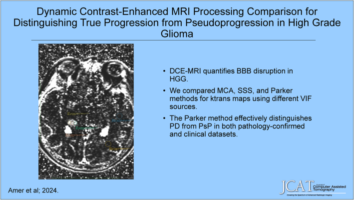
Dynamic Contrast-enhanced MRI Processing Comparison for Distinguishing True Progression From Pseudoprogression in High-grade Glioma.
Background: Treatment-related changes may occur due to radiation and temozolomide in glioblastoma and can mimic tumor progression on conventional MRI. DCE-MRI enables quantification of the extent of blood-brain barrier (BBB) disruption, providing information about areas of suspicious postcontrast T1 enhancement. We compared DCE-MRI processing methods for distinguishing true disease progression from pseudoprogression in high-grade gliomas (HGGs).
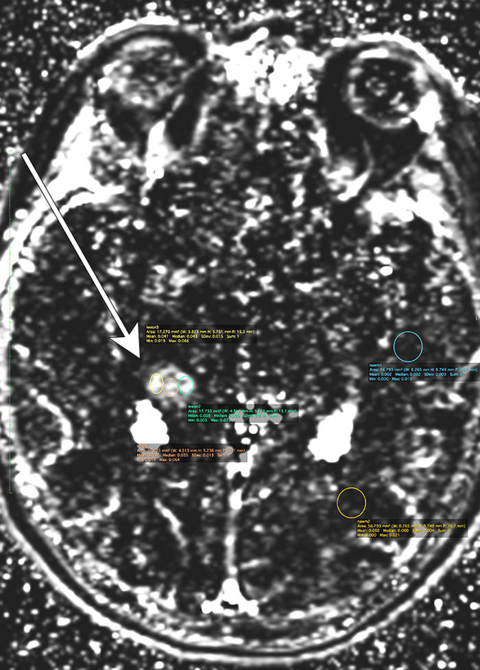
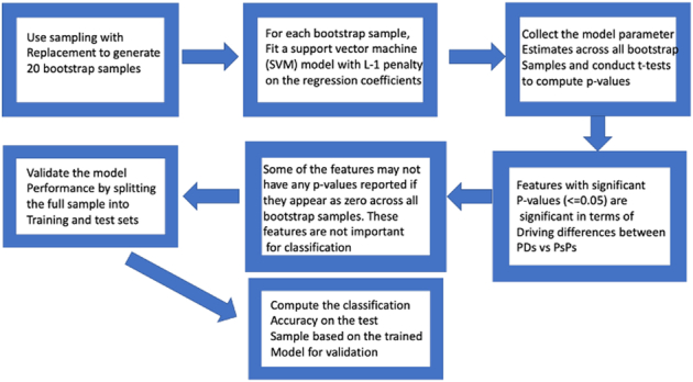
Methods: We identified 110 patients with HGG treated with surgery and chemoradiation who underwent DCE-MRI to further interrogate areas of new/increasing enhancement. All patients had confirmatory surgery/biopsy with pathology-confirmed progression or pseudoprogression. Scans were performed at 3T and analyzed using nordicICE. The MCA, SSS, and Parker models are three standardized processing methodologies used to create k trans maps, a parameter that quantifies BBB permeability. Three equal regions of interest were placed at sites of peak contrast enhancement within each lesion. Data from each method was processed for mean and maximum k trans . We conducted several rounds of analysis and finalized a strategy on penalized support vector machines based on engineered features with bootstrap sampling.
Results: The Parker method was significant for k trans maximum in the combined pathology and clinical as well as the pathology-only data sets. MCA and SSS did not perform well under the SVM classifier for pathology only. For clinical follow-up subjects, the Parker method yielded statistically significant results for maximum and mean k trans .
Conclusions: The Parker method was effective in distinguishing PD and PsP for pathology and clinical data sets. MCA and SSS techniques were effective for the clinical data set.
期刊介绍:
The mission of Journal of Computer Assisted Tomography is to showcase the latest clinical and research developments in CT, MR, and closely related diagnostic techniques. We encourage submission of both original research and review articles that have immediate or promissory clinical applications. Topics of special interest include: 1) functional MR and CT of the brain and body; 2) advanced/innovative MRI techniques (diffusion, perfusion, rapid scanning); and 3) advanced/innovative CT techniques (perfusion, multi-energy, dose-reduction, and processing).

 求助内容:
求助内容: 应助结果提醒方式:


