Parkin过表达可促进线粒体自噬对运动性中暑大鼠肺的保护作用。
引用次数: 0
摘要
背景:Pink1/Parkin通路和线粒体自噬在中暑肺损伤中的作用尚不清楚。本研究探讨了Pink1/ parkinson介导的线粒体自噬在运动性中暑(EHS)大鼠急性肺损伤(ALI)中的作用。方法:将60只sd大鼠随机分为对照组(CON)、对照组+ Parkin过表达组(CON + Parkin)、EHS组和EHS + Parkin过表达组(EHS + Parkin)。通过向尾静脉注射携带Parkin基因的腺相关病毒,过表达Parkin,建立EHS大鼠模型。采用微计算机断层扫描(micro-CT)分析肺组织病理变化,测定肺系数和肺毛细血管通透性。采用酶联免疫吸附法测定白细胞介素-6 (IL-6)、IL-1β、肿瘤坏死因子-α和活性氧水平。透射电镜观察肺组织Ⅱ型上皮细胞线粒体形态;免疫荧光法检测肺组织凋亡、线粒体自噬水平以及Pink1和Parkin的共定位。采用Western blotting检测大鼠肺组织中Pink1、Parkin、mitofusin-2 (MFN2)、磷酸酶和紧张素同源物(PTEN)、PTEN- l、p62及自噬标志物微管相关蛋白1轻链3 (LC3)的表达,并计算LC3II/LC3I的比值。结果:与EHS组比较,EHS + Parkin组大鼠存活率明显高于EHS组。大鼠肺系数和肺血管通透性降低,病理改变明显减轻(P P P >0.05)。结论:Pink1/Parkin介导的线粒体自噬功能障碍是EHS大鼠ALI的机制之一,激活Parkin过表达介导的线粒体自噬可减轻EHS引起的ALI。本文章由计算机程序翻译,如有差异,请以英文原文为准。
Overexpression of Parkin promotes the protective effect of mitochondrial autophagy on the lung of rats with exertional heatstroke
Background
The roles of the Pink1/Parkin pathway and mitophagy in lung injury during heat stroke remain unclear. In this study, we investigated the role of Pink1/Parkin-mediated mitophagy in acute lung injury (ALI) in rats with exertional heat stroke (EHS).
Methods
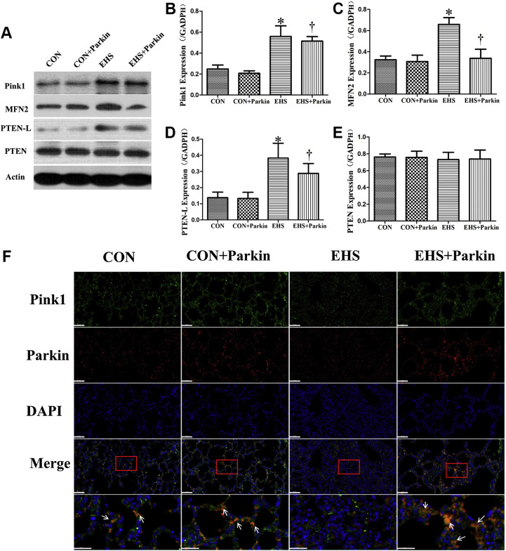
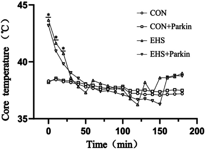
Sixty Sprague Dawley rats were randomly divided into control (CON), control + Parkin overexpression (CON + Parkin), EHS, and EHS + Parkin overexpression (EHS + Parkin) groups. Parkin was overexpressed by injecting an adeno-associated virus carrying the Parkin gene into the tail vein, and a rat model of EHS was established. Pathological changes in the lung tissue were analyzed using microcomputed tomography (micro-CT), and the lung coefficient and pulmonary capillary permeability were measured. Enzyme-linked immunosorbent assay were used to determine the levels of interleukin-6 (IL-6), IL-1β, and tumor necrosis factor-α, and reactive oxygen species. The morphology of mitochondria in type Ⅱ epithelial cells of lung tissue was observed using transmission electron microscopy; and the apoptosis of lung tissue, the level of mitophagy, and the co-localization of Pink1 and Parkin were determined using immunofluorescence. The expression of Pink1, Parkin, mitofusin-2 (MFN2), phosphatase and tensin homolog (PTEN), PTEN-L, p62, and the autophagy marker microtubule-associated protein 1 light chain 3 (LC3) in rat lung tissue was measured by Western blotting, and the ratio of LC3II/LC3I was calculated.
Results
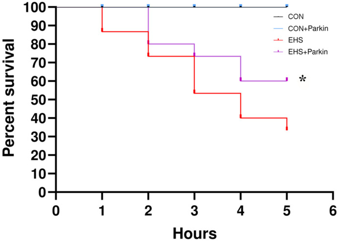
Compared with the EHS group, the survival rate of rats in the EHS + Parkin group was significantly higher. Their lung coefficient and pulmonary vascular permeability decreased and the pathological changes were significantly alleviated (P <0.05). Their levels of inflammatory factors and reactive oxygen species were significantly decreased (P <0.05), and the degree of mitochondrial swelling in pulmonary type II epithelial cells was alleviated. The apoptosis of lung tissue was alleviated, the colocalization of Pink1 and Parkin, LC3 and Tom20 was enhanced, and the ratio of LC3-II/LC3-I increased. The expression of Pink1, MFN2, PTEN-L, and p62 decreased, whereas the expression of PTEN was not significantly different from that in the EHS group (P >0.05).
Conclusion
Pink1/Parkin-mediated mitophagy dysfunction is one of the mechanisms underlying ALI in rats with EHS, and activation of Parkin overexpression-mediated mitophagy can alleviate ALI caused by EHS.
求助全文
通过发布文献求助,成功后即可免费获取论文全文。
去求助
来源期刊

Journal of intensive medicine
Critical Care and Intensive Care Medicine
CiteScore
1.90
自引率
0.00%
发文量
0
审稿时长
58 days
 求助内容:
求助内容: 应助结果提醒方式:


