Ning Chai, Tanja Stachon, Sabrina Häcker, Tim Berger, Zhen Li, Maryam Amini, Shweta Suiwal, Berthold Seitz, Achim Langenbucher, Nóra Szentmáry
{"title":"脂多糖诱导的角膜人类角膜成纤维细胞炎症对 RB-PDT 的细胞反应:细胞因子、趋化因子及相关信号通路的启示。","authors":"Ning Chai, Tanja Stachon, Sabrina Häcker, Tim Berger, Zhen Li, Maryam Amini, Shweta Suiwal, Berthold Seitz, Achim Langenbucher, Nóra Szentmáry","doi":"10.1371/journal.pone.0318132","DOIUrl":null,"url":null,"abstract":"<p><strong>Purpose: </strong>Rose Bengal Photodynamic Therapy (RB-PDT) offers dual therapeutic benefits by enhancing corneal stiffness and providing antibacterial activity, presenting significant potential for patients with keratoconus complicated by keratitis. Our purpose was to assess the effect of rose bengal photodynamic therapy (RB-PDT) on the expression of pro-inflammatory cytokines and chemokines, as well as on extracellular matrix (ECM)-related molecules, in lipopolysaccharide (LPS)-induced inflammation of keratoconus human corneal fibroblasts (KC-HCFs). Additionally, the involvement of the mitogen-activated protein kinase (MAPK) and nuclear factor kappa B (NF-κB) signaling pathways which are downstream of the Toll-like receptor 4 (TLR4) pathway were examined.</p><p><strong>Methods: </strong>KC-HCFs were stimulated with varying concentrations of LPS (0-10 μg/ml), which was followed by RB-PDT. The expression levels of interleukin-1β (IL-1β), IL-6, IL-8, interferon alpha 2 (IFNA2), IFNB1, intercellular adhesion molecule 1 (ICAM-1), chemokine (C-C motif) ligand 4 (CCL-4), collagen I, collagen V, lysyl oxidase (LOX), transforming growth factor β 1(TGF-β1) were measured using qPCR, ELISA, or western blot. The activation of the NF-κB and MAPK pathways was assessed using qPCR and western blot.</p><p><strong>Results: </strong>In LPS-induced inflammation of KC-HCFs, the expression of IL-6 was further amplified by the treatment with RB-PDT (p = 0.001). However, the activation of the MAPK and NF-κB pathways did not increase following RB-PDT. Additionally, RB-PDT reduced the transcription of collagen I and collagen V (p≤0.03), while the transcription of LOX and TGF-β1 secretion remained unchanged in KC-HCFs exposed to LPS.</p><p><strong>Conclusion: </strong>In LPS-induced inflammation of KC-HCFs treated with RB-PDT, despite the increased expression of pro-inflammatory cytokines, the activation of the TLR4 signaling pathways is lacking. RB-PDT may have no adverse effects on corneal scar formation of keratoconus corneas in the short term.</p>","PeriodicalId":20189,"journal":{"name":"PLoS ONE","volume":"20 1","pages":"e0318132"},"PeriodicalIF":2.6000,"publicationDate":"2025-01-27","publicationTypes":"Journal Article","fieldsOfStudy":null,"isOpenAccess":false,"openAccessPdf":"https://www.ncbi.nlm.nih.gov/pmc/articles/PMC11771863/pdf/","citationCount":"0","resultStr":"{\"title\":\"The cellular response of lipopolysaccharide-induced inflammation in keratoconus human corneal fibroblasts to RB-PDT: Insights into cytokines, chemokines and related signaling pathways.\",\"authors\":\"Ning Chai, Tanja Stachon, Sabrina Häcker, Tim Berger, Zhen Li, Maryam Amini, Shweta Suiwal, Berthold Seitz, Achim Langenbucher, Nóra Szentmáry\",\"doi\":\"10.1371/journal.pone.0318132\",\"DOIUrl\":null,\"url\":null,\"abstract\":\"<p><strong>Purpose: </strong>Rose Bengal Photodynamic Therapy (RB-PDT) offers dual therapeutic benefits by enhancing corneal stiffness and providing antibacterial activity, presenting significant potential for patients with keratoconus complicated by keratitis. Our purpose was to assess the effect of rose bengal photodynamic therapy (RB-PDT) on the expression of pro-inflammatory cytokines and chemokines, as well as on extracellular matrix (ECM)-related molecules, in lipopolysaccharide (LPS)-induced inflammation of keratoconus human corneal fibroblasts (KC-HCFs). Additionally, the involvement of the mitogen-activated protein kinase (MAPK) and nuclear factor kappa B (NF-κB) signaling pathways which are downstream of the Toll-like receptor 4 (TLR4) pathway were examined.</p><p><strong>Methods: </strong>KC-HCFs were stimulated with varying concentrations of LPS (0-10 μg/ml), which was followed by RB-PDT. The expression levels of interleukin-1β (IL-1β), IL-6, IL-8, interferon alpha 2 (IFNA2), IFNB1, intercellular adhesion molecule 1 (ICAM-1), chemokine (C-C motif) ligand 4 (CCL-4), collagen I, collagen V, lysyl oxidase (LOX), transforming growth factor β 1(TGF-β1) were measured using qPCR, ELISA, or western blot. The activation of the NF-κB and MAPK pathways was assessed using qPCR and western blot.</p><p><strong>Results: </strong>In LPS-induced inflammation of KC-HCFs, the expression of IL-6 was further amplified by the treatment with RB-PDT (p = 0.001). However, the activation of the MAPK and NF-κB pathways did not increase following RB-PDT. Additionally, RB-PDT reduced the transcription of collagen I and collagen V (p≤0.03), while the transcription of LOX and TGF-β1 secretion remained unchanged in KC-HCFs exposed to LPS.</p><p><strong>Conclusion: </strong>In LPS-induced inflammation of KC-HCFs treated with RB-PDT, despite the increased expression of pro-inflammatory cytokines, the activation of the TLR4 signaling pathways is lacking. RB-PDT may have no adverse effects on corneal scar formation of keratoconus corneas in the short term.</p>\",\"PeriodicalId\":20189,\"journal\":{\"name\":\"PLoS ONE\",\"volume\":\"20 1\",\"pages\":\"e0318132\"},\"PeriodicalIF\":2.6000,\"publicationDate\":\"2025-01-27\",\"publicationTypes\":\"Journal Article\",\"fieldsOfStudy\":null,\"isOpenAccess\":false,\"openAccessPdf\":\"https://www.ncbi.nlm.nih.gov/pmc/articles/PMC11771863/pdf/\",\"citationCount\":\"0\",\"resultStr\":null,\"platform\":\"Semanticscholar\",\"paperid\":null,\"PeriodicalName\":\"PLoS ONE\",\"FirstCategoryId\":\"103\",\"ListUrlMain\":\"https://doi.org/10.1371/journal.pone.0318132\",\"RegionNum\":3,\"RegionCategory\":\"综合性期刊\",\"ArticlePicture\":[],\"TitleCN\":null,\"AbstractTextCN\":null,\"PMCID\":null,\"EPubDate\":\"2025/1/1 0:00:00\",\"PubModel\":\"eCollection\",\"JCR\":\"Q1\",\"JCRName\":\"MULTIDISCIPLINARY SCIENCES\",\"Score\":null,\"Total\":0}","platform":"Semanticscholar","paperid":null,"PeriodicalName":"PLoS ONE","FirstCategoryId":"103","ListUrlMain":"https://doi.org/10.1371/journal.pone.0318132","RegionNum":3,"RegionCategory":"综合性期刊","ArticlePicture":[],"TitleCN":null,"AbstractTextCN":null,"PMCID":null,"EPubDate":"2025/1/1 0:00:00","PubModel":"eCollection","JCR":"Q1","JCRName":"MULTIDISCIPLINARY SCIENCES","Score":null,"Total":0}
The cellular response of lipopolysaccharide-induced inflammation in keratoconus human corneal fibroblasts to RB-PDT: Insights into cytokines, chemokines and related signaling pathways.
Purpose: Rose Bengal Photodynamic Therapy (RB-PDT) offers dual therapeutic benefits by enhancing corneal stiffness and providing antibacterial activity, presenting significant potential for patients with keratoconus complicated by keratitis. Our purpose was to assess the effect of rose bengal photodynamic therapy (RB-PDT) on the expression of pro-inflammatory cytokines and chemokines, as well as on extracellular matrix (ECM)-related molecules, in lipopolysaccharide (LPS)-induced inflammation of keratoconus human corneal fibroblasts (KC-HCFs). Additionally, the involvement of the mitogen-activated protein kinase (MAPK) and nuclear factor kappa B (NF-κB) signaling pathways which are downstream of the Toll-like receptor 4 (TLR4) pathway were examined.
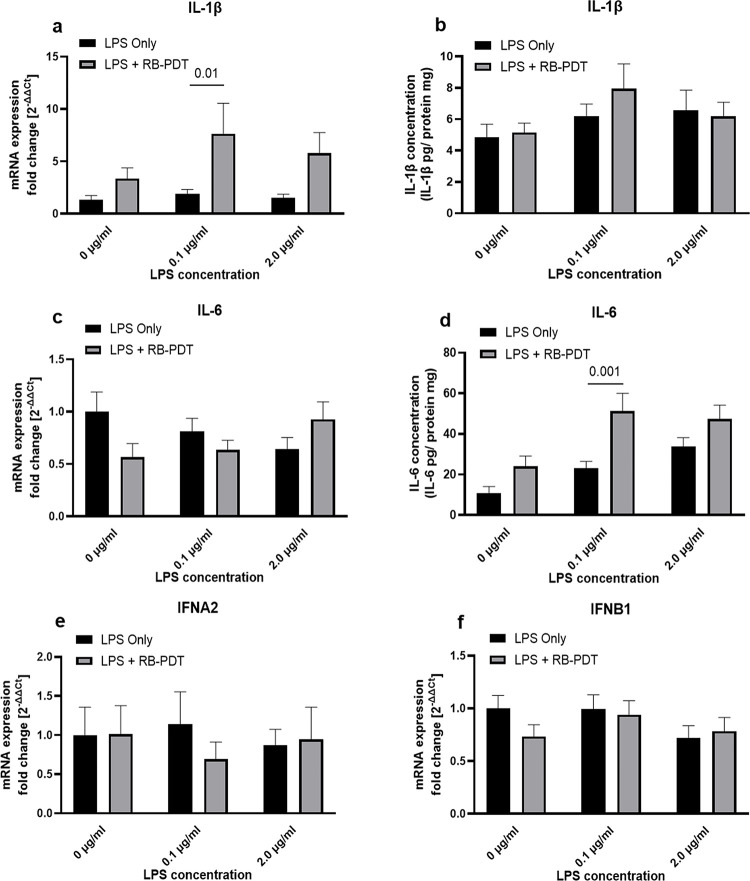
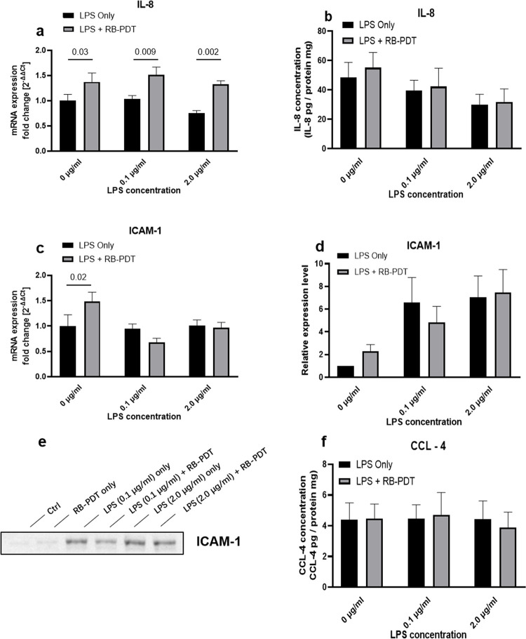
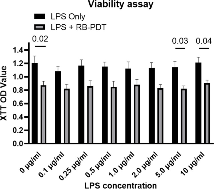
Methods: KC-HCFs were stimulated with varying concentrations of LPS (0-10 μg/ml), which was followed by RB-PDT. The expression levels of interleukin-1β (IL-1β), IL-6, IL-8, interferon alpha 2 (IFNA2), IFNB1, intercellular adhesion molecule 1 (ICAM-1), chemokine (C-C motif) ligand 4 (CCL-4), collagen I, collagen V, lysyl oxidase (LOX), transforming growth factor β 1(TGF-β1) were measured using qPCR, ELISA, or western blot. The activation of the NF-κB and MAPK pathways was assessed using qPCR and western blot.
Results: In LPS-induced inflammation of KC-HCFs, the expression of IL-6 was further amplified by the treatment with RB-PDT (p = 0.001). However, the activation of the MAPK and NF-κB pathways did not increase following RB-PDT. Additionally, RB-PDT reduced the transcription of collagen I and collagen V (p≤0.03), while the transcription of LOX and TGF-β1 secretion remained unchanged in KC-HCFs exposed to LPS.
Conclusion: In LPS-induced inflammation of KC-HCFs treated with RB-PDT, despite the increased expression of pro-inflammatory cytokines, the activation of the TLR4 signaling pathways is lacking. RB-PDT may have no adverse effects on corneal scar formation of keratoconus corneas in the short term.
期刊介绍:
PLOS ONE is an international, peer-reviewed, open-access, online publication. PLOS ONE welcomes reports on primary research from any scientific discipline. It provides:
* Open-access—freely accessible online, authors retain copyright
* Fast publication times
* Peer review by expert, practicing researchers
* Post-publication tools to indicate quality and impact
* Community-based dialogue on articles
* Worldwide media coverage

 求助内容:
求助内容: 应助结果提醒方式:


