Cinzia Santucciu, Ashkan Hajjafari, Soheil Sadr, Scilla Mastrandrea, Carlo Rettaroli, Luca Simbula, Mariano Scaglione, Salvatore Masala, Angela Peruzzu, Giovanna Masala
{"title":"罕见的胆固醇晶体形成在一个罕见的临床病例报告的脾包虫病囊肿的病人从撒丁岛,意大利。","authors":"Cinzia Santucciu, Ashkan Hajjafari, Soheil Sadr, Scilla Mastrandrea, Carlo Rettaroli, Luca Simbula, Mariano Scaglione, Salvatore Masala, Angela Peruzzu, Giovanna Masala","doi":"10.3389/fpara.2024.1498099","DOIUrl":null,"url":null,"abstract":"<p><p>Cystic echinococcosis (CE) is a zoonotic disease caused by <i>Echinococcus granulosus</i> sensu lato, the metacestode of a tapeworm parasite of high medical importance. Infection of the parasite leads to the development of echinococcal cysts, and the spleen is a rarely infected organ. A 46-year-old woman who was born and who resides in Sardinia, Italy, was referred to the Echinococcosis outpatient clinic at the University Hospital of Sassari (Sardinia, Italy) for a pain in the left flank. She used to live in the countryside, in contact with several animals, and for 2 years, she had been working in a family garden, growing vegetables as a hobby. Ultrasounds and X-ray were performed, which evidenced a rounded formation in the upper third of the spleen, while a CT scan confirmed a parasitological cyst. Immunological examinations on serum samples did not detect specific antibodies against <i>Echinococcus</i> spp. Following surgical exportation, the whole spleen with the cystic lesion was delivered to the World Organisation for Animal Health (WOAH) and the National Reference Laboratory for Echinococcosis for further laboratory analyses. Moreover, characterization of the cyst fluid resulted dense and shiny. Observation under a light microscope at ×400 magnification revealed the formation of rectangular crystals and aggregates attributable to cholesterol molecules. Subsequently, through parasitological investigation, molecular biology investigations confirmed <i>E. granulosus</i> sensu stricto G1. We describe cholesterol crystals in a splenic echinococcal cyst for the first time. There is no clear explanation of how the cholesterol crystals formed in this case, but this was attributed to multifactorial causes, including atherosclerosis, chronic inflammation, parasite metabolism, and host responses.</p>","PeriodicalId":73098,"journal":{"name":"Frontiers in parasitology","volume":"3 ","pages":"1498099"},"PeriodicalIF":0.0000,"publicationDate":"2025-01-10","publicationTypes":"Journal Article","fieldsOfStudy":null,"isOpenAccess":false,"openAccessPdf":"https://www.ncbi.nlm.nih.gov/pmc/articles/PMC11759282/pdf/","citationCount":"0","resultStr":"{\"title\":\"Unusual cholesterol crystal formation in a rare clinical case report of splenic echinococcal cyst in a patient from Sardinia, Italy.\",\"authors\":\"Cinzia Santucciu, Ashkan Hajjafari, Soheil Sadr, Scilla Mastrandrea, Carlo Rettaroli, Luca Simbula, Mariano Scaglione, Salvatore Masala, Angela Peruzzu, Giovanna Masala\",\"doi\":\"10.3389/fpara.2024.1498099\",\"DOIUrl\":null,\"url\":null,\"abstract\":\"<p><p>Cystic echinococcosis (CE) is a zoonotic disease caused by <i>Echinococcus granulosus</i> sensu lato, the metacestode of a tapeworm parasite of high medical importance. Infection of the parasite leads to the development of echinococcal cysts, and the spleen is a rarely infected organ. A 46-year-old woman who was born and who resides in Sardinia, Italy, was referred to the Echinococcosis outpatient clinic at the University Hospital of Sassari (Sardinia, Italy) for a pain in the left flank. She used to live in the countryside, in contact with several animals, and for 2 years, she had been working in a family garden, growing vegetables as a hobby. Ultrasounds and X-ray were performed, which evidenced a rounded formation in the upper third of the spleen, while a CT scan confirmed a parasitological cyst. Immunological examinations on serum samples did not detect specific antibodies against <i>Echinococcus</i> spp. Following surgical exportation, the whole spleen with the cystic lesion was delivered to the World Organisation for Animal Health (WOAH) and the National Reference Laboratory for Echinococcosis for further laboratory analyses. Moreover, characterization of the cyst fluid resulted dense and shiny. Observation under a light microscope at ×400 magnification revealed the formation of rectangular crystals and aggregates attributable to cholesterol molecules. Subsequently, through parasitological investigation, molecular biology investigations confirmed <i>E. granulosus</i> sensu stricto G1. We describe cholesterol crystals in a splenic echinococcal cyst for the first time. There is no clear explanation of how the cholesterol crystals formed in this case, but this was attributed to multifactorial causes, including atherosclerosis, chronic inflammation, parasite metabolism, and host responses.</p>\",\"PeriodicalId\":73098,\"journal\":{\"name\":\"Frontiers in parasitology\",\"volume\":\"3 \",\"pages\":\"1498099\"},\"PeriodicalIF\":0.0000,\"publicationDate\":\"2025-01-10\",\"publicationTypes\":\"Journal Article\",\"fieldsOfStudy\":null,\"isOpenAccess\":false,\"openAccessPdf\":\"https://www.ncbi.nlm.nih.gov/pmc/articles/PMC11759282/pdf/\",\"citationCount\":\"0\",\"resultStr\":null,\"platform\":\"Semanticscholar\",\"paperid\":null,\"PeriodicalName\":\"Frontiers in parasitology\",\"FirstCategoryId\":\"1085\",\"ListUrlMain\":\"https://doi.org/10.3389/fpara.2024.1498099\",\"RegionNum\":0,\"RegionCategory\":null,\"ArticlePicture\":[],\"TitleCN\":null,\"AbstractTextCN\":null,\"PMCID\":null,\"EPubDate\":\"2024/1/1 0:00:00\",\"PubModel\":\"eCollection\",\"JCR\":\"\",\"JCRName\":\"\",\"Score\":null,\"Total\":0}","platform":"Semanticscholar","paperid":null,"PeriodicalName":"Frontiers in parasitology","FirstCategoryId":"1085","ListUrlMain":"https://doi.org/10.3389/fpara.2024.1498099","RegionNum":0,"RegionCategory":null,"ArticlePicture":[],"TitleCN":null,"AbstractTextCN":null,"PMCID":null,"EPubDate":"2024/1/1 0:00:00","PubModel":"eCollection","JCR":"","JCRName":"","Score":null,"Total":0}
Unusual cholesterol crystal formation in a rare clinical case report of splenic echinococcal cyst in a patient from Sardinia, Italy.
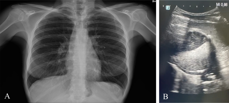
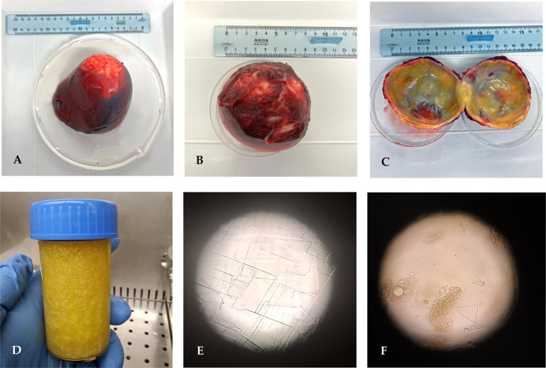
Cystic echinococcosis (CE) is a zoonotic disease caused by Echinococcus granulosus sensu lato, the metacestode of a tapeworm parasite of high medical importance. Infection of the parasite leads to the development of echinococcal cysts, and the spleen is a rarely infected organ. A 46-year-old woman who was born and who resides in Sardinia, Italy, was referred to the Echinococcosis outpatient clinic at the University Hospital of Sassari (Sardinia, Italy) for a pain in the left flank. She used to live in the countryside, in contact with several animals, and for 2 years, she had been working in a family garden, growing vegetables as a hobby. Ultrasounds and X-ray were performed, which evidenced a rounded formation in the upper third of the spleen, while a CT scan confirmed a parasitological cyst. Immunological examinations on serum samples did not detect specific antibodies against Echinococcus spp. Following surgical exportation, the whole spleen with the cystic lesion was delivered to the World Organisation for Animal Health (WOAH) and the National Reference Laboratory for Echinococcosis for further laboratory analyses. Moreover, characterization of the cyst fluid resulted dense and shiny. Observation under a light microscope at ×400 magnification revealed the formation of rectangular crystals and aggregates attributable to cholesterol molecules. Subsequently, through parasitological investigation, molecular biology investigations confirmed E. granulosus sensu stricto G1. We describe cholesterol crystals in a splenic echinococcal cyst for the first time. There is no clear explanation of how the cholesterol crystals formed in this case, but this was attributed to multifactorial causes, including atherosclerosis, chronic inflammation, parasite metabolism, and host responses.

 求助内容:
求助内容: 应助结果提醒方式:


