{"title":"胆管消失综合征中巨噬细胞和肝细胞S1PR2/RhoA/ROCK1/MLC2通路上S1PR2的激活","authors":"Aya Miyagawa-Hayashino, Tetsuya Imura, Toshiaki Takezawa, Maki Hirai, Saya Shibata, Hiroshi Ogi, Takahiro Tsujikawa, Eiichi Konishi","doi":"10.1371/journal.pone.0317568","DOIUrl":null,"url":null,"abstract":"<p><p>Immunologic bile duct destruction is a pathogenic condition associated with vanishing bile duct syndrome (VBDS) after liver transplantation and hematopoietic stem-cell transplantation. As the bile acid receptor sphingosine 1-phosphate receptor 2 (S1PR2) plays a critical role in recruitment of bone marrow-derived monocytes/macrophages to sites of cholestatic liver injury, S1PR2 expression was examined using cultured macrophages and patient tissues. Bile canaliculi destruction precedes intrahepatic ductopenia; therefore, we focused on hepatocyte S1PR2 and the downstream RhoA/Rho kinase 1 (ROCK1) signaling pathway and bile canaliculi alterations using three-dimensional hepatocyte culture models that form obvious bile canaliculus-like networks. Multiplex immunohistochemistry revealed increased numbers of S1PR2+CD45+CD68+FCN1+ inflammatory macrophages and S1PR2+CD45+CD68+MARCO+ Kupffer cells in liver tissues showing ductopenia due to graft-versus-host disease and rejection post-liver transplant compared with normal liver. Macrophage expression of proinflammatory cytokines, including MCP1, was reduced following S1PR2 inhibition. Taurocholic acid and S1P2 agonist induced hepatocyte S1PR2 and reduced RhoA/ROCK1 expression, resulting in bile canaliculi dilatation. S1PR2 inhibition reversed the effect on RhoA/ROCK1 expression, resulting in maintenance of bile canaliculi through myosin light chain 2 (MLC2) phosphorylation. Activation of S1PR2 on macrophages and S1PR2 on hepatocytes may disrupt bile canaliculi dynamics in VBDS under regulation by RhoA/ROCK1 through MLC2 phosphorylation.</p>","PeriodicalId":20189,"journal":{"name":"PLoS ONE","volume":"20 1","pages":"e0317568"},"PeriodicalIF":2.6000,"publicationDate":"2025-01-24","publicationTypes":"Journal Article","fieldsOfStudy":null,"isOpenAccess":false,"openAccessPdf":"https://www.ncbi.nlm.nih.gov/pmc/articles/PMC11760576/pdf/","citationCount":"0","resultStr":"{\"title\":\"Activation of S1PR2 on macrophages and the hepatocyte S1PR2/RhoA/ROCK1/MLC2 pathway in vanishing bile duct syndrome.\",\"authors\":\"Aya Miyagawa-Hayashino, Tetsuya Imura, Toshiaki Takezawa, Maki Hirai, Saya Shibata, Hiroshi Ogi, Takahiro Tsujikawa, Eiichi Konishi\",\"doi\":\"10.1371/journal.pone.0317568\",\"DOIUrl\":null,\"url\":null,\"abstract\":\"<p><p>Immunologic bile duct destruction is a pathogenic condition associated with vanishing bile duct syndrome (VBDS) after liver transplantation and hematopoietic stem-cell transplantation. As the bile acid receptor sphingosine 1-phosphate receptor 2 (S1PR2) plays a critical role in recruitment of bone marrow-derived monocytes/macrophages to sites of cholestatic liver injury, S1PR2 expression was examined using cultured macrophages and patient tissues. Bile canaliculi destruction precedes intrahepatic ductopenia; therefore, we focused on hepatocyte S1PR2 and the downstream RhoA/Rho kinase 1 (ROCK1) signaling pathway and bile canaliculi alterations using three-dimensional hepatocyte culture models that form obvious bile canaliculus-like networks. Multiplex immunohistochemistry revealed increased numbers of S1PR2+CD45+CD68+FCN1+ inflammatory macrophages and S1PR2+CD45+CD68+MARCO+ Kupffer cells in liver tissues showing ductopenia due to graft-versus-host disease and rejection post-liver transplant compared with normal liver. Macrophage expression of proinflammatory cytokines, including MCP1, was reduced following S1PR2 inhibition. Taurocholic acid and S1P2 agonist induced hepatocyte S1PR2 and reduced RhoA/ROCK1 expression, resulting in bile canaliculi dilatation. S1PR2 inhibition reversed the effect on RhoA/ROCK1 expression, resulting in maintenance of bile canaliculi through myosin light chain 2 (MLC2) phosphorylation. Activation of S1PR2 on macrophages and S1PR2 on hepatocytes may disrupt bile canaliculi dynamics in VBDS under regulation by RhoA/ROCK1 through MLC2 phosphorylation.</p>\",\"PeriodicalId\":20189,\"journal\":{\"name\":\"PLoS ONE\",\"volume\":\"20 1\",\"pages\":\"e0317568\"},\"PeriodicalIF\":2.6000,\"publicationDate\":\"2025-01-24\",\"publicationTypes\":\"Journal Article\",\"fieldsOfStudy\":null,\"isOpenAccess\":false,\"openAccessPdf\":\"https://www.ncbi.nlm.nih.gov/pmc/articles/PMC11760576/pdf/\",\"citationCount\":\"0\",\"resultStr\":null,\"platform\":\"Semanticscholar\",\"paperid\":null,\"PeriodicalName\":\"PLoS ONE\",\"FirstCategoryId\":\"103\",\"ListUrlMain\":\"https://doi.org/10.1371/journal.pone.0317568\",\"RegionNum\":3,\"RegionCategory\":\"综合性期刊\",\"ArticlePicture\":[],\"TitleCN\":null,\"AbstractTextCN\":null,\"PMCID\":null,\"EPubDate\":\"2025/1/1 0:00:00\",\"PubModel\":\"eCollection\",\"JCR\":\"Q1\",\"JCRName\":\"MULTIDISCIPLINARY SCIENCES\",\"Score\":null,\"Total\":0}","platform":"Semanticscholar","paperid":null,"PeriodicalName":"PLoS ONE","FirstCategoryId":"103","ListUrlMain":"https://doi.org/10.1371/journal.pone.0317568","RegionNum":3,"RegionCategory":"综合性期刊","ArticlePicture":[],"TitleCN":null,"AbstractTextCN":null,"PMCID":null,"EPubDate":"2025/1/1 0:00:00","PubModel":"eCollection","JCR":"Q1","JCRName":"MULTIDISCIPLINARY SCIENCES","Score":null,"Total":0}
Activation of S1PR2 on macrophages and the hepatocyte S1PR2/RhoA/ROCK1/MLC2 pathway in vanishing bile duct syndrome.
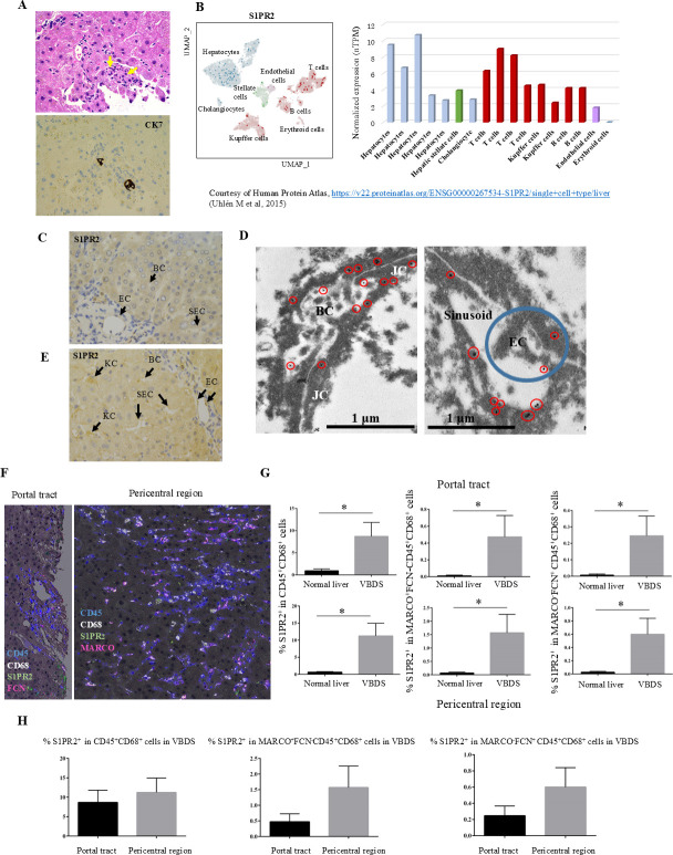
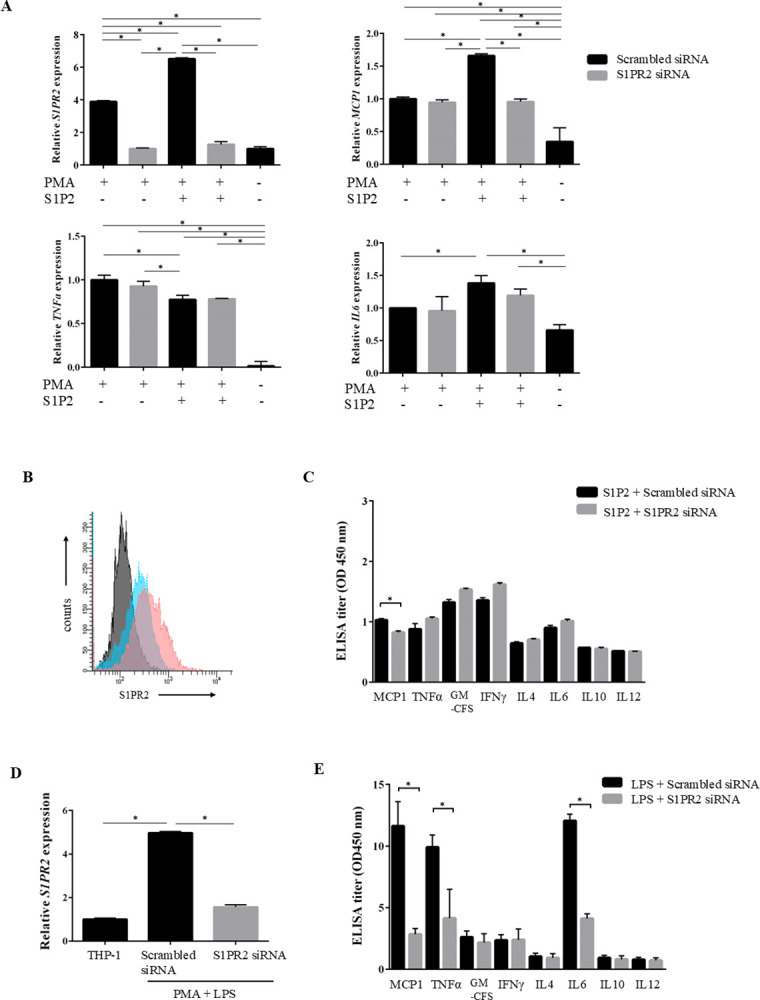
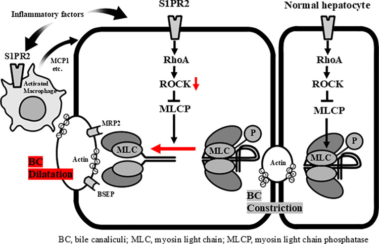
Immunologic bile duct destruction is a pathogenic condition associated with vanishing bile duct syndrome (VBDS) after liver transplantation and hematopoietic stem-cell transplantation. As the bile acid receptor sphingosine 1-phosphate receptor 2 (S1PR2) plays a critical role in recruitment of bone marrow-derived monocytes/macrophages to sites of cholestatic liver injury, S1PR2 expression was examined using cultured macrophages and patient tissues. Bile canaliculi destruction precedes intrahepatic ductopenia; therefore, we focused on hepatocyte S1PR2 and the downstream RhoA/Rho kinase 1 (ROCK1) signaling pathway and bile canaliculi alterations using three-dimensional hepatocyte culture models that form obvious bile canaliculus-like networks. Multiplex immunohistochemistry revealed increased numbers of S1PR2+CD45+CD68+FCN1+ inflammatory macrophages and S1PR2+CD45+CD68+MARCO+ Kupffer cells in liver tissues showing ductopenia due to graft-versus-host disease and rejection post-liver transplant compared with normal liver. Macrophage expression of proinflammatory cytokines, including MCP1, was reduced following S1PR2 inhibition. Taurocholic acid and S1P2 agonist induced hepatocyte S1PR2 and reduced RhoA/ROCK1 expression, resulting in bile canaliculi dilatation. S1PR2 inhibition reversed the effect on RhoA/ROCK1 expression, resulting in maintenance of bile canaliculi through myosin light chain 2 (MLC2) phosphorylation. Activation of S1PR2 on macrophages and S1PR2 on hepatocytes may disrupt bile canaliculi dynamics in VBDS under regulation by RhoA/ROCK1 through MLC2 phosphorylation.
期刊介绍:
PLOS ONE is an international, peer-reviewed, open-access, online publication. PLOS ONE welcomes reports on primary research from any scientific discipline. It provides:
* Open-access—freely accessible online, authors retain copyright
* Fast publication times
* Peer review by expert, practicing researchers
* Post-publication tools to indicate quality and impact
* Community-based dialogue on articles
* Worldwide media coverage

 求助内容:
求助内容: 应助结果提醒方式:


