Burak Durmaz, Latife Merve Oktay Çelebi, Ayşe Çekin, Ayshan Ahadova, Nur Selvi Günel, Hatice Kalkan Yıldırım, Ali Mert Özgönül, Eser Yıldırım Sözmen
{"title":"蜂胶对PPP2R1A及癌细胞凋亡的影响","authors":"Burak Durmaz, Latife Merve Oktay Çelebi, Ayşe Çekin, Ayshan Ahadova, Nur Selvi Günel, Hatice Kalkan Yıldırım, Ali Mert Özgönül, Eser Yıldırım Sözmen","doi":"10.1155/bri/5538068","DOIUrl":null,"url":null,"abstract":"<p><p>Recently, it has been shown that protein phosphatase 2A (PP2A) dysfunction was common in many cancer types and was mediated by various inactivation mechanisms. Although many research studies observed antitumor effect of propolis extracts in various types of cancer, the mechanism of effect are still obscure. In this study, we investigated the effect of propolis on PPP2R1A expression and its relationship with apoptosis in the SW-620 (colorectal cancer), DU-145 and PC-3 (prostate cancer), and MCF-7 (breast cancer) cell lines, with WI-38 (healthy fibroblast) cells serving as the control. Moreover, we aimed to investigate the impact of propolis on apoptosis by analyzing apoptosis markers such as tumor necrosis factor-related apoptosis-inducing ligand (TRAIL), APAF-1, and caspases-3, -8, and -9. Propolis samples were extracted, and their phenolic compounds were quantified using LC-MS/MS. The RealTime Cell Analysis System-xCELLigence (RTCA-SP) device and software were employed to assess cell viability and cytotoxicity of the propolis samples. The IC<sub>50</sub> values for propolis were determined (298 μg/mL for SW-620, 185.6 μg/mL for DU-145, 250.7 μg/mL for PC - 3, 292.9 μg/mL for MCF-7, and 311.2 μg/mL for WI-38). Subsequently, the effects of propolis on PPP2R1A expression and apoptosis markers (TRAIL, Apaf-1, and caspases-3, -8, and -9) were analyzed. When we compared the healthy cell lines to cancer cell lines, a statistically significant increase in caspase-3 (3.62-fold) and in TRAIL (4.38-fold) was observed in the SW-620 cell line after the application of propolis. In addition, in the PC-3 cell line, a 1.4-fold increase in caspase-8 was observed compared with the healthy cell line, which is also statistically significant. Our findings indicated that propolis increased the PPP2R1A levels and apoptosis markers in cancer cell lines. It has been suggested that high PPP2R1A levels induced by propolis treatment might activate the apoptosis pathway. In this study, the inducible effect of propolis on PPP2R1A levels, identified as a new target for cancer treatment, was demonstrated for the first time. The findings suggest that propolis holds promise as a potential cancer therapy by increasing PPP2R1A levels, a key molecule in cancer treatment.</p>","PeriodicalId":8826,"journal":{"name":"Biochemistry Research International","volume":"2025 ","pages":"5538068"},"PeriodicalIF":3.4000,"publicationDate":"2025-01-15","publicationTypes":"Journal Article","fieldsOfStudy":null,"isOpenAccess":false,"openAccessPdf":"https://www.ncbi.nlm.nih.gov/pmc/articles/PMC11756940/pdf/","citationCount":"0","resultStr":"{\"title\":\"Effect of Propolis on PPP2R1A and Apoptosis in Cancer Cells.\",\"authors\":\"Burak Durmaz, Latife Merve Oktay Çelebi, Ayşe Çekin, Ayshan Ahadova, Nur Selvi Günel, Hatice Kalkan Yıldırım, Ali Mert Özgönül, Eser Yıldırım Sözmen\",\"doi\":\"10.1155/bri/5538068\",\"DOIUrl\":null,\"url\":null,\"abstract\":\"<p><p>Recently, it has been shown that protein phosphatase 2A (PP2A) dysfunction was common in many cancer types and was mediated by various inactivation mechanisms. Although many research studies observed antitumor effect of propolis extracts in various types of cancer, the mechanism of effect are still obscure. In this study, we investigated the effect of propolis on PPP2R1A expression and its relationship with apoptosis in the SW-620 (colorectal cancer), DU-145 and PC-3 (prostate cancer), and MCF-7 (breast cancer) cell lines, with WI-38 (healthy fibroblast) cells serving as the control. Moreover, we aimed to investigate the impact of propolis on apoptosis by analyzing apoptosis markers such as tumor necrosis factor-related apoptosis-inducing ligand (TRAIL), APAF-1, and caspases-3, -8, and -9. Propolis samples were extracted, and their phenolic compounds were quantified using LC-MS/MS. The RealTime Cell Analysis System-xCELLigence (RTCA-SP) device and software were employed to assess cell viability and cytotoxicity of the propolis samples. The IC<sub>50</sub> values for propolis were determined (298 μg/mL for SW-620, 185.6 μg/mL for DU-145, 250.7 μg/mL for PC - 3, 292.9 μg/mL for MCF-7, and 311.2 μg/mL for WI-38). Subsequently, the effects of propolis on PPP2R1A expression and apoptosis markers (TRAIL, Apaf-1, and caspases-3, -8, and -9) were analyzed. When we compared the healthy cell lines to cancer cell lines, a statistically significant increase in caspase-3 (3.62-fold) and in TRAIL (4.38-fold) was observed in the SW-620 cell line after the application of propolis. In addition, in the PC-3 cell line, a 1.4-fold increase in caspase-8 was observed compared with the healthy cell line, which is also statistically significant. Our findings indicated that propolis increased the PPP2R1A levels and apoptosis markers in cancer cell lines. It has been suggested that high PPP2R1A levels induced by propolis treatment might activate the apoptosis pathway. In this study, the inducible effect of propolis on PPP2R1A levels, identified as a new target for cancer treatment, was demonstrated for the first time. The findings suggest that propolis holds promise as a potential cancer therapy by increasing PPP2R1A levels, a key molecule in cancer treatment.</p>\",\"PeriodicalId\":8826,\"journal\":{\"name\":\"Biochemistry Research International\",\"volume\":\"2025 \",\"pages\":\"5538068\"},\"PeriodicalIF\":3.4000,\"publicationDate\":\"2025-01-15\",\"publicationTypes\":\"Journal Article\",\"fieldsOfStudy\":null,\"isOpenAccess\":false,\"openAccessPdf\":\"https://www.ncbi.nlm.nih.gov/pmc/articles/PMC11756940/pdf/\",\"citationCount\":\"0\",\"resultStr\":null,\"platform\":\"Semanticscholar\",\"paperid\":null,\"PeriodicalName\":\"Biochemistry Research International\",\"FirstCategoryId\":\"1085\",\"ListUrlMain\":\"https://doi.org/10.1155/bri/5538068\",\"RegionNum\":0,\"RegionCategory\":null,\"ArticlePicture\":[],\"TitleCN\":null,\"AbstractTextCN\":null,\"PMCID\":null,\"EPubDate\":\"2025/1/1 0:00:00\",\"PubModel\":\"eCollection\",\"JCR\":\"Q2\",\"JCRName\":\"BIOCHEMICAL RESEARCH METHODS\",\"Score\":null,\"Total\":0}","platform":"Semanticscholar","paperid":null,"PeriodicalName":"Biochemistry Research International","FirstCategoryId":"1085","ListUrlMain":"https://doi.org/10.1155/bri/5538068","RegionNum":0,"RegionCategory":null,"ArticlePicture":[],"TitleCN":null,"AbstractTextCN":null,"PMCID":null,"EPubDate":"2025/1/1 0:00:00","PubModel":"eCollection","JCR":"Q2","JCRName":"BIOCHEMICAL RESEARCH METHODS","Score":null,"Total":0}
Effect of Propolis on PPP2R1A and Apoptosis in Cancer Cells.
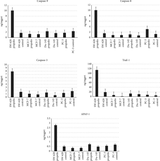
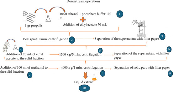
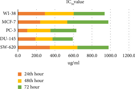
Recently, it has been shown that protein phosphatase 2A (PP2A) dysfunction was common in many cancer types and was mediated by various inactivation mechanisms. Although many research studies observed antitumor effect of propolis extracts in various types of cancer, the mechanism of effect are still obscure. In this study, we investigated the effect of propolis on PPP2R1A expression and its relationship with apoptosis in the SW-620 (colorectal cancer), DU-145 and PC-3 (prostate cancer), and MCF-7 (breast cancer) cell lines, with WI-38 (healthy fibroblast) cells serving as the control. Moreover, we aimed to investigate the impact of propolis on apoptosis by analyzing apoptosis markers such as tumor necrosis factor-related apoptosis-inducing ligand (TRAIL), APAF-1, and caspases-3, -8, and -9. Propolis samples were extracted, and their phenolic compounds were quantified using LC-MS/MS. The RealTime Cell Analysis System-xCELLigence (RTCA-SP) device and software were employed to assess cell viability and cytotoxicity of the propolis samples. The IC50 values for propolis were determined (298 μg/mL for SW-620, 185.6 μg/mL for DU-145, 250.7 μg/mL for PC - 3, 292.9 μg/mL for MCF-7, and 311.2 μg/mL for WI-38). Subsequently, the effects of propolis on PPP2R1A expression and apoptosis markers (TRAIL, Apaf-1, and caspases-3, -8, and -9) were analyzed. When we compared the healthy cell lines to cancer cell lines, a statistically significant increase in caspase-3 (3.62-fold) and in TRAIL (4.38-fold) was observed in the SW-620 cell line after the application of propolis. In addition, in the PC-3 cell line, a 1.4-fold increase in caspase-8 was observed compared with the healthy cell line, which is also statistically significant. Our findings indicated that propolis increased the PPP2R1A levels and apoptosis markers in cancer cell lines. It has been suggested that high PPP2R1A levels induced by propolis treatment might activate the apoptosis pathway. In this study, the inducible effect of propolis on PPP2R1A levels, identified as a new target for cancer treatment, was demonstrated for the first time. The findings suggest that propolis holds promise as a potential cancer therapy by increasing PPP2R1A levels, a key molecule in cancer treatment.

 求助内容:
求助内容: 应助结果提醒方式:


