{"title":"人类腰椎3D打印模拟物的开发和生物力学评估。","authors":"Siril Teja Dukkipati, Mark Driscoll","doi":"10.1186/s41205-025-00249-y","DOIUrl":null,"url":null,"abstract":"<p><strong>Background: </strong>There exists a need for validated lumbar spine models in spine biomechanics research. Although cadaveric testing is the current gold standard for spinal implant development, it poses significant issues related to reliability and repeatability due to the wide variability in cadaveric physiologies. Moreover, there are increasing ethical concerns with human dissection practices. Analogue models can act as cost saving alternatives to human tissue with better repeatability. The current study proposes a new methodology of spinal biomechanics testing using 3D printable surrogates and characterized its multi-dimensional stiffness in displacement-controlled loading scenarios.</p><p><strong>Methods: </strong>The model consisted of L1 to S1 vertebrae, intervertebral discs (IVD), intertransverse, interspinous, anterior and posterior longitudinal ligaments. The vertebrae and the IVDs were derived from an open-source 3D MRI anatomography database, while the ligaments were modeled based on literature incorporating mounting points on the spinous and transverse processes. Stereolithography 3D printing along with a combination of stiff and soft photopolymer resins were used to manufacture the vertebrae and the soft tissues in the model. Thereafter, displacement-controlled pure moments were applied in the range of ± 15° at 0.5°/sec in all bending modes using a torsion testing machine and a custom pure bending jig. Model rotation and resisting moment under loading were recorded to quantify the rotational stiffness and hysteresis in the model.</p><p><strong>Results: </strong>The model reached a maximum of 5.66Nm and 3.53Nm at 15° flexion-extension, 3.84Nm and 3.93Nm at 15° right and left lateral bending, and 2.45Nm and 2.59Nm at 15° right and left axial rotation respectively. Model RMS error against ex vivo human response was estimated to be 1.57°, 1.64°, 0.82° in flexion-extension, lateral bending and axial rotation respectively. Bilateral symmetry in model stiffness was observed in lateral bending and axial rotation directions.</p><p><strong>Conclusions: </strong>This study presents a reproducible 3D printable L1-S1 lumbar spine and validated it in all three orthogonal bending modes in the range of ± 15° against ex vivo and in silico data. The 3D printed analogue spine model described herein shows promising results, suggesting this model, with further validation, could have potential as a human cadaveric tissue substitute within the explored contexts of use.</p>","PeriodicalId":72036,"journal":{"name":"3D printing in medicine","volume":"11 1","pages":"3"},"PeriodicalIF":3.1000,"publicationDate":"2025-01-23","publicationTypes":"Journal Article","fieldsOfStudy":null,"isOpenAccess":false,"openAccessPdf":"https://www.ncbi.nlm.nih.gov/pmc/articles/PMC11755917/pdf/","citationCount":"0","resultStr":"{\"title\":\"Development and biomechanical evaluation of a 3D printed analogue of the human lumbar spine.\",\"authors\":\"Siril Teja Dukkipati, Mark Driscoll\",\"doi\":\"10.1186/s41205-025-00249-y\",\"DOIUrl\":null,\"url\":null,\"abstract\":\"<p><strong>Background: </strong>There exists a need for validated lumbar spine models in spine biomechanics research. Although cadaveric testing is the current gold standard for spinal implant development, it poses significant issues related to reliability and repeatability due to the wide variability in cadaveric physiologies. Moreover, there are increasing ethical concerns with human dissection practices. Analogue models can act as cost saving alternatives to human tissue with better repeatability. The current study proposes a new methodology of spinal biomechanics testing using 3D printable surrogates and characterized its multi-dimensional stiffness in displacement-controlled loading scenarios.</p><p><strong>Methods: </strong>The model consisted of L1 to S1 vertebrae, intervertebral discs (IVD), intertransverse, interspinous, anterior and posterior longitudinal ligaments. The vertebrae and the IVDs were derived from an open-source 3D MRI anatomography database, while the ligaments were modeled based on literature incorporating mounting points on the spinous and transverse processes. Stereolithography 3D printing along with a combination of stiff and soft photopolymer resins were used to manufacture the vertebrae and the soft tissues in the model. Thereafter, displacement-controlled pure moments were applied in the range of ± 15° at 0.5°/sec in all bending modes using a torsion testing machine and a custom pure bending jig. Model rotation and resisting moment under loading were recorded to quantify the rotational stiffness and hysteresis in the model.</p><p><strong>Results: </strong>The model reached a maximum of 5.66Nm and 3.53Nm at 15° flexion-extension, 3.84Nm and 3.93Nm at 15° right and left lateral bending, and 2.45Nm and 2.59Nm at 15° right and left axial rotation respectively. Model RMS error against ex vivo human response was estimated to be 1.57°, 1.64°, 0.82° in flexion-extension, lateral bending and axial rotation respectively. Bilateral symmetry in model stiffness was observed in lateral bending and axial rotation directions.</p><p><strong>Conclusions: </strong>This study presents a reproducible 3D printable L1-S1 lumbar spine and validated it in all three orthogonal bending modes in the range of ± 15° against ex vivo and in silico data. The 3D printed analogue spine model described herein shows promising results, suggesting this model, with further validation, could have potential as a human cadaveric tissue substitute within the explored contexts of use.</p>\",\"PeriodicalId\":72036,\"journal\":{\"name\":\"3D printing in medicine\",\"volume\":\"11 1\",\"pages\":\"3\"},\"PeriodicalIF\":3.1000,\"publicationDate\":\"2025-01-23\",\"publicationTypes\":\"Journal Article\",\"fieldsOfStudy\":null,\"isOpenAccess\":false,\"openAccessPdf\":\"https://www.ncbi.nlm.nih.gov/pmc/articles/PMC11755917/pdf/\",\"citationCount\":\"0\",\"resultStr\":null,\"platform\":\"Semanticscholar\",\"paperid\":null,\"PeriodicalName\":\"3D printing in medicine\",\"FirstCategoryId\":\"1085\",\"ListUrlMain\":\"https://doi.org/10.1186/s41205-025-00249-y\",\"RegionNum\":0,\"RegionCategory\":null,\"ArticlePicture\":[],\"TitleCN\":null,\"AbstractTextCN\":null,\"PMCID\":null,\"EPubDate\":\"\",\"PubModel\":\"\",\"JCR\":\"Q1\",\"JCRName\":\"RADIOLOGY, NUCLEAR MEDICINE & MEDICAL IMAGING\",\"Score\":null,\"Total\":0}","platform":"Semanticscholar","paperid":null,"PeriodicalName":"3D printing in medicine","FirstCategoryId":"1085","ListUrlMain":"https://doi.org/10.1186/s41205-025-00249-y","RegionNum":0,"RegionCategory":null,"ArticlePicture":[],"TitleCN":null,"AbstractTextCN":null,"PMCID":null,"EPubDate":"","PubModel":"","JCR":"Q1","JCRName":"RADIOLOGY, NUCLEAR MEDICINE & MEDICAL IMAGING","Score":null,"Total":0}
Development and biomechanical evaluation of a 3D printed analogue of the human lumbar spine.
Background: There exists a need for validated lumbar spine models in spine biomechanics research. Although cadaveric testing is the current gold standard for spinal implant development, it poses significant issues related to reliability and repeatability due to the wide variability in cadaveric physiologies. Moreover, there are increasing ethical concerns with human dissection practices. Analogue models can act as cost saving alternatives to human tissue with better repeatability. The current study proposes a new methodology of spinal biomechanics testing using 3D printable surrogates and characterized its multi-dimensional stiffness in displacement-controlled loading scenarios.
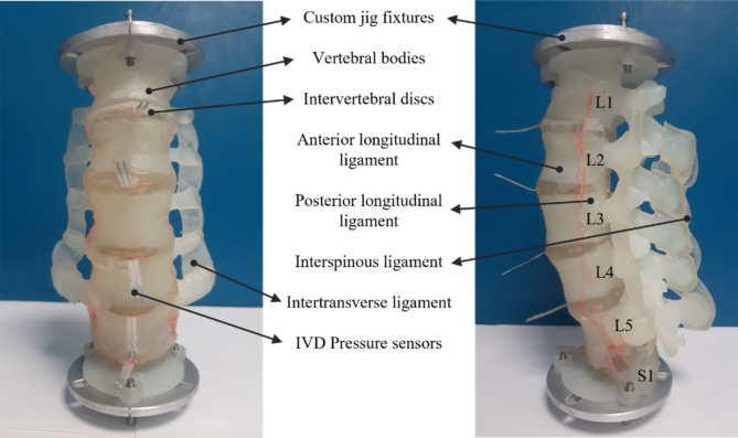
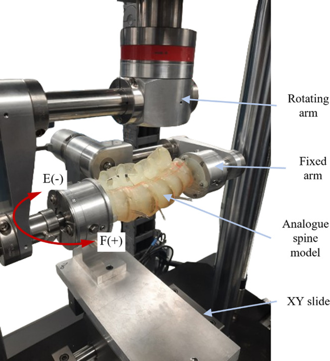
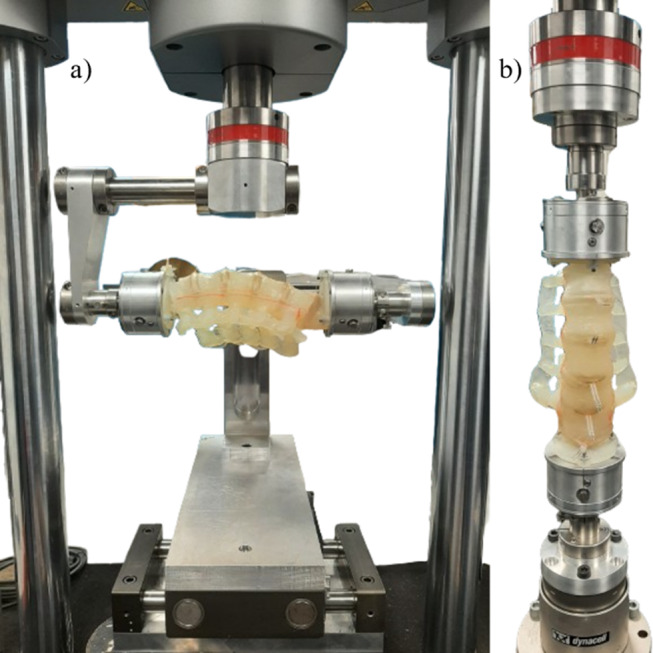
Methods: The model consisted of L1 to S1 vertebrae, intervertebral discs (IVD), intertransverse, interspinous, anterior and posterior longitudinal ligaments. The vertebrae and the IVDs were derived from an open-source 3D MRI anatomography database, while the ligaments were modeled based on literature incorporating mounting points on the spinous and transverse processes. Stereolithography 3D printing along with a combination of stiff and soft photopolymer resins were used to manufacture the vertebrae and the soft tissues in the model. Thereafter, displacement-controlled pure moments were applied in the range of ± 15° at 0.5°/sec in all bending modes using a torsion testing machine and a custom pure bending jig. Model rotation and resisting moment under loading were recorded to quantify the rotational stiffness and hysteresis in the model.
Results: The model reached a maximum of 5.66Nm and 3.53Nm at 15° flexion-extension, 3.84Nm and 3.93Nm at 15° right and left lateral bending, and 2.45Nm and 2.59Nm at 15° right and left axial rotation respectively. Model RMS error against ex vivo human response was estimated to be 1.57°, 1.64°, 0.82° in flexion-extension, lateral bending and axial rotation respectively. Bilateral symmetry in model stiffness was observed in lateral bending and axial rotation directions.
Conclusions: This study presents a reproducible 3D printable L1-S1 lumbar spine and validated it in all three orthogonal bending modes in the range of ± 15° against ex vivo and in silico data. The 3D printed analogue spine model described herein shows promising results, suggesting this model, with further validation, could have potential as a human cadaveric tissue substitute within the explored contexts of use.

 求助内容:
求助内容: 应助结果提醒方式:


