Erika Mayumi Watanabe, Ronan Yudi Cavazzana, Douglas de Aguiar Manso Ribeiro, Lorena Candido Brandão, Ana Victória Haddad, José Eduardo Corrente, André Petean Trindade, Eliane Chaves Jorge
{"title":"Graves眼病眼外肌和眼球突出的计算机断层形态学分析。","authors":"Erika Mayumi Watanabe, Ronan Yudi Cavazzana, Douglas de Aguiar Manso Ribeiro, Lorena Candido Brandão, Ana Victória Haddad, José Eduardo Corrente, André Petean Trindade, Eliane Chaves Jorge","doi":"10.1590/0100-3984.2024.0040-en","DOIUrl":null,"url":null,"abstract":"<p><strong>Objective: </strong>To assess the prevalence of changes on computed tomography (CT) in Graves' orbitopathy (GO) and to correlate those changes with disease activity, as well as with clinical and biochemical variables.</p><p><strong>Materials and methods: </strong>This was a retrospective study, conducted at a tertiary hospital, of clinical, biochemical, and imaging data from consecutive patients with GO who underwent at least one orbital CT scan between July 2012 and December 2020. A single observer quantified the thickness of the extraocular muscles and the degree of proptosis. Clinical and biochemical variables were analyzed to determine whether they correlated with CT changes, GO activity, and GO severity.</p><p><strong>Results: </strong>Our sample included data from 67 patients with GO (134 orbits), 50 (74.6%) of whom were female. There were positive correlations between the clinical activity score and increase in thyroid-stimulating factor/free thyroxine, between the severity of GO and the increase in the thickness of the extraocular muscles, and between the degree of proptosis and muscle thickness.</p><p><strong>Conclusion: </strong>Orbital CT proved effective in detecting thickening of the extraocular muscles and proptosis in patients with GO, changes that correlated significantly with clinical and biochemical variables. Muscle thickening was associated with the severity of GO and could be a biomarker of the risk of vision loss.</p>","PeriodicalId":20842,"journal":{"name":"Radiologia Brasileira","volume":"57 ","pages":"e20240040en"},"PeriodicalIF":0.0000,"publicationDate":"2024-12-27","publicationTypes":"Journal Article","fieldsOfStudy":null,"isOpenAccess":false,"openAccessPdf":"https://www.ncbi.nlm.nih.gov/pmc/articles/PMC11734998/pdf/","citationCount":"0","resultStr":"{\"title\":\"Morphometric analysis of extraocular muscles and proptosis by computed tomography in Graves' orbitopathy.\",\"authors\":\"Erika Mayumi Watanabe, Ronan Yudi Cavazzana, Douglas de Aguiar Manso Ribeiro, Lorena Candido Brandão, Ana Victória Haddad, José Eduardo Corrente, André Petean Trindade, Eliane Chaves Jorge\",\"doi\":\"10.1590/0100-3984.2024.0040-en\",\"DOIUrl\":null,\"url\":null,\"abstract\":\"<p><strong>Objective: </strong>To assess the prevalence of changes on computed tomography (CT) in Graves' orbitopathy (GO) and to correlate those changes with disease activity, as well as with clinical and biochemical variables.</p><p><strong>Materials and methods: </strong>This was a retrospective study, conducted at a tertiary hospital, of clinical, biochemical, and imaging data from consecutive patients with GO who underwent at least one orbital CT scan between July 2012 and December 2020. A single observer quantified the thickness of the extraocular muscles and the degree of proptosis. Clinical and biochemical variables were analyzed to determine whether they correlated with CT changes, GO activity, and GO severity.</p><p><strong>Results: </strong>Our sample included data from 67 patients with GO (134 orbits), 50 (74.6%) of whom were female. There were positive correlations between the clinical activity score and increase in thyroid-stimulating factor/free thyroxine, between the severity of GO and the increase in the thickness of the extraocular muscles, and between the degree of proptosis and muscle thickness.</p><p><strong>Conclusion: </strong>Orbital CT proved effective in detecting thickening of the extraocular muscles and proptosis in patients with GO, changes that correlated significantly with clinical and biochemical variables. Muscle thickening was associated with the severity of GO and could be a biomarker of the risk of vision loss.</p>\",\"PeriodicalId\":20842,\"journal\":{\"name\":\"Radiologia Brasileira\",\"volume\":\"57 \",\"pages\":\"e20240040en\"},\"PeriodicalIF\":0.0000,\"publicationDate\":\"2024-12-27\",\"publicationTypes\":\"Journal Article\",\"fieldsOfStudy\":null,\"isOpenAccess\":false,\"openAccessPdf\":\"https://www.ncbi.nlm.nih.gov/pmc/articles/PMC11734998/pdf/\",\"citationCount\":\"0\",\"resultStr\":null,\"platform\":\"Semanticscholar\",\"paperid\":null,\"PeriodicalName\":\"Radiologia Brasileira\",\"FirstCategoryId\":\"1085\",\"ListUrlMain\":\"https://doi.org/10.1590/0100-3984.2024.0040-en\",\"RegionNum\":0,\"RegionCategory\":null,\"ArticlePicture\":[],\"TitleCN\":null,\"AbstractTextCN\":null,\"PMCID\":null,\"EPubDate\":\"2024/1/1 0:00:00\",\"PubModel\":\"eCollection\",\"JCR\":\"Q3\",\"JCRName\":\"Medicine\",\"Score\":null,\"Total\":0}","platform":"Semanticscholar","paperid":null,"PeriodicalName":"Radiologia Brasileira","FirstCategoryId":"1085","ListUrlMain":"https://doi.org/10.1590/0100-3984.2024.0040-en","RegionNum":0,"RegionCategory":null,"ArticlePicture":[],"TitleCN":null,"AbstractTextCN":null,"PMCID":null,"EPubDate":"2024/1/1 0:00:00","PubModel":"eCollection","JCR":"Q3","JCRName":"Medicine","Score":null,"Total":0}
Morphometric analysis of extraocular muscles and proptosis by computed tomography in Graves' orbitopathy.
Objective: To assess the prevalence of changes on computed tomography (CT) in Graves' orbitopathy (GO) and to correlate those changes with disease activity, as well as with clinical and biochemical variables.
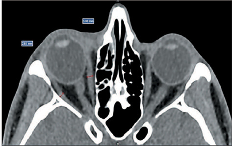
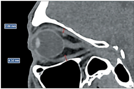
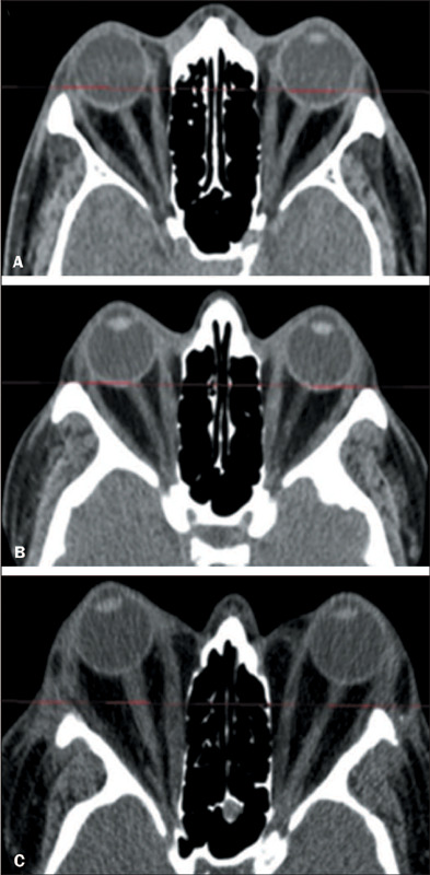
Materials and methods: This was a retrospective study, conducted at a tertiary hospital, of clinical, biochemical, and imaging data from consecutive patients with GO who underwent at least one orbital CT scan between July 2012 and December 2020. A single observer quantified the thickness of the extraocular muscles and the degree of proptosis. Clinical and biochemical variables were analyzed to determine whether they correlated with CT changes, GO activity, and GO severity.
Results: Our sample included data from 67 patients with GO (134 orbits), 50 (74.6%) of whom were female. There were positive correlations between the clinical activity score and increase in thyroid-stimulating factor/free thyroxine, between the severity of GO and the increase in the thickness of the extraocular muscles, and between the degree of proptosis and muscle thickness.
Conclusion: Orbital CT proved effective in detecting thickening of the extraocular muscles and proptosis in patients with GO, changes that correlated significantly with clinical and biochemical variables. Muscle thickening was associated with the severity of GO and could be a biomarker of the risk of vision loss.

 求助内容:
求助内容: 应助结果提醒方式:


