{"title":"当比较共聚焦激光扫描和光学切片显微镜获得的小胶质细胞形态学特征时,观察到最小的差异。","authors":"Sânziana Godeanu, Mădălina Iuliana Mușat, Anja Scheller, Eugen Osiac, Bogdan Cătălin","doi":"10.3389/fnana.2024.1507140","DOIUrl":null,"url":null,"abstract":"<p><strong>Background: </strong>While widefield microscopy has long been constrained by out-of-focus scattering, advancements have generated a solution in the form of confocal laser scanning microscopy (cLSM) and optical sectioning microscopy using structured illumination (OSM). In this study, we aim to investigate, using microglia branching, if cLSM and OSM can produce images with comparable morphological characteristics.</p><p><strong>Results: </strong>By imaging the somatosensory microglia from a tissue slice of a 3-week-old mouse and establishing morphological parameters that characterizes the microglial branching pattern, we were able to show that there is no difference in total length of the branch tree, number of branches, mean branch length and number of primary to terminal branches. We did find that area-based parameters such as mean occupied area and mean surveillance area were bigger in cLSM isolated microglia compared to OSM ones. Additionally, by investigating the difference in acquisition time between techniques and personal costs we were able to establish that the amortization could be made in 6.11 ± 2.93 years in the case of countries with a Human Development Index (HDI) = 7-9 and 7.06 ± 3.13 years, respectably, for countries with HDI < 7. As such, OSM systems seem a valid option if one just wants basic histological evaluation, and cLSM should be considered for groups that demand higher resolution or volumetric images.</p>","PeriodicalId":12572,"journal":{"name":"Frontiers in Neuroanatomy","volume":"18 ","pages":"1507140"},"PeriodicalIF":2.3000,"publicationDate":"2025-01-03","publicationTypes":"Journal Article","fieldsOfStudy":null,"isOpenAccess":false,"openAccessPdf":"https://www.ncbi.nlm.nih.gov/pmc/articles/PMC11739110/pdf/","citationCount":"0","resultStr":"{\"title\":\"Minimal differences observed when comparing the morphological profiling of microglia obtained by confocal laser scanning and optical sectioning microscopy.\",\"authors\":\"Sânziana Godeanu, Mădălina Iuliana Mușat, Anja Scheller, Eugen Osiac, Bogdan Cătălin\",\"doi\":\"10.3389/fnana.2024.1507140\",\"DOIUrl\":null,\"url\":null,\"abstract\":\"<p><strong>Background: </strong>While widefield microscopy has long been constrained by out-of-focus scattering, advancements have generated a solution in the form of confocal laser scanning microscopy (cLSM) and optical sectioning microscopy using structured illumination (OSM). In this study, we aim to investigate, using microglia branching, if cLSM and OSM can produce images with comparable morphological characteristics.</p><p><strong>Results: </strong>By imaging the somatosensory microglia from a tissue slice of a 3-week-old mouse and establishing morphological parameters that characterizes the microglial branching pattern, we were able to show that there is no difference in total length of the branch tree, number of branches, mean branch length and number of primary to terminal branches. We did find that area-based parameters such as mean occupied area and mean surveillance area were bigger in cLSM isolated microglia compared to OSM ones. Additionally, by investigating the difference in acquisition time between techniques and personal costs we were able to establish that the amortization could be made in 6.11 ± 2.93 years in the case of countries with a Human Development Index (HDI) = 7-9 and 7.06 ± 3.13 years, respectably, for countries with HDI < 7. As such, OSM systems seem a valid option if one just wants basic histological evaluation, and cLSM should be considered for groups that demand higher resolution or volumetric images.</p>\",\"PeriodicalId\":12572,\"journal\":{\"name\":\"Frontiers in Neuroanatomy\",\"volume\":\"18 \",\"pages\":\"1507140\"},\"PeriodicalIF\":2.3000,\"publicationDate\":\"2025-01-03\",\"publicationTypes\":\"Journal Article\",\"fieldsOfStudy\":null,\"isOpenAccess\":false,\"openAccessPdf\":\"https://www.ncbi.nlm.nih.gov/pmc/articles/PMC11739110/pdf/\",\"citationCount\":\"0\",\"resultStr\":null,\"platform\":\"Semanticscholar\",\"paperid\":null,\"PeriodicalName\":\"Frontiers in Neuroanatomy\",\"FirstCategoryId\":\"3\",\"ListUrlMain\":\"https://doi.org/10.3389/fnana.2024.1507140\",\"RegionNum\":4,\"RegionCategory\":\"医学\",\"ArticlePicture\":[],\"TitleCN\":null,\"AbstractTextCN\":null,\"PMCID\":null,\"EPubDate\":\"2024/1/1 0:00:00\",\"PubModel\":\"eCollection\",\"JCR\":\"Q1\",\"JCRName\":\"ANATOMY & MORPHOLOGY\",\"Score\":null,\"Total\":0}","platform":"Semanticscholar","paperid":null,"PeriodicalName":"Frontiers in Neuroanatomy","FirstCategoryId":"3","ListUrlMain":"https://doi.org/10.3389/fnana.2024.1507140","RegionNum":4,"RegionCategory":"医学","ArticlePicture":[],"TitleCN":null,"AbstractTextCN":null,"PMCID":null,"EPubDate":"2024/1/1 0:00:00","PubModel":"eCollection","JCR":"Q1","JCRName":"ANATOMY & MORPHOLOGY","Score":null,"Total":0}
Minimal differences observed when comparing the morphological profiling of microglia obtained by confocal laser scanning and optical sectioning microscopy.
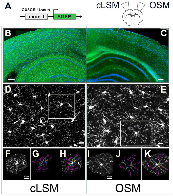
Background: While widefield microscopy has long been constrained by out-of-focus scattering, advancements have generated a solution in the form of confocal laser scanning microscopy (cLSM) and optical sectioning microscopy using structured illumination (OSM). In this study, we aim to investigate, using microglia branching, if cLSM and OSM can produce images with comparable morphological characteristics.
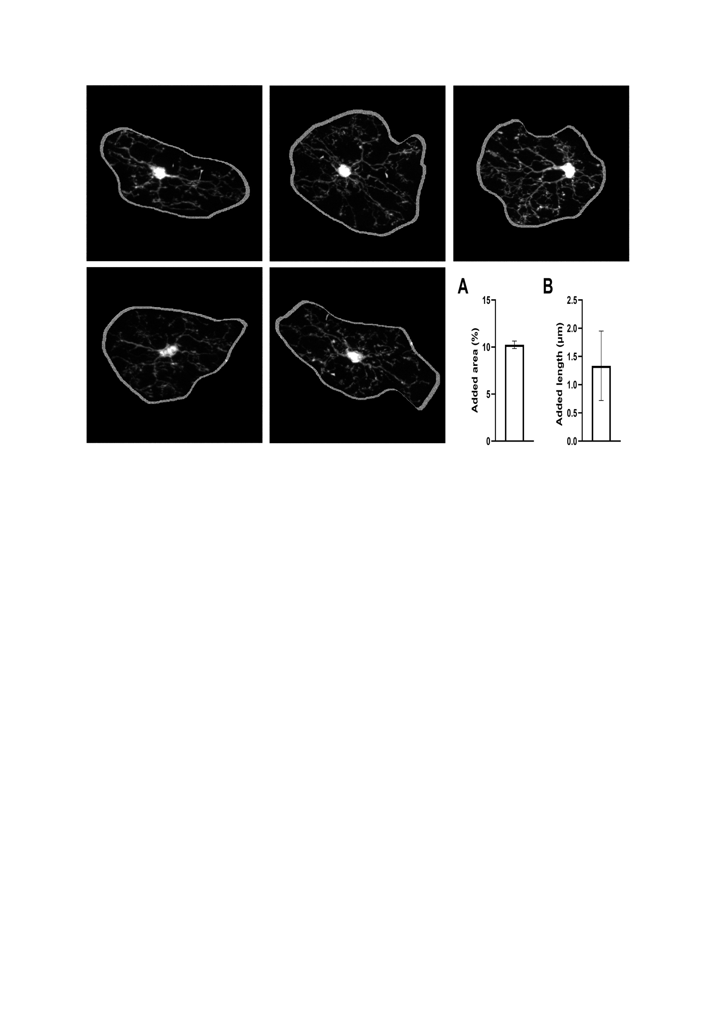
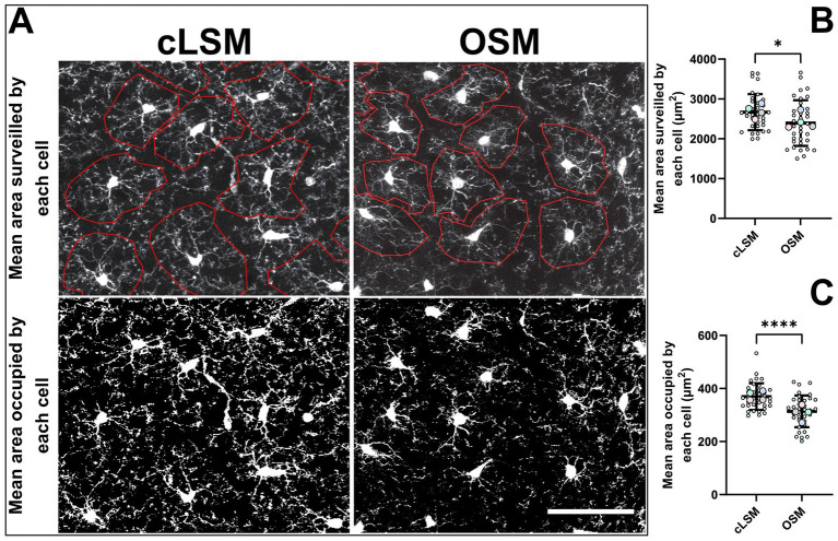
Results: By imaging the somatosensory microglia from a tissue slice of a 3-week-old mouse and establishing morphological parameters that characterizes the microglial branching pattern, we were able to show that there is no difference in total length of the branch tree, number of branches, mean branch length and number of primary to terminal branches. We did find that area-based parameters such as mean occupied area and mean surveillance area were bigger in cLSM isolated microglia compared to OSM ones. Additionally, by investigating the difference in acquisition time between techniques and personal costs we were able to establish that the amortization could be made in 6.11 ± 2.93 years in the case of countries with a Human Development Index (HDI) = 7-9 and 7.06 ± 3.13 years, respectably, for countries with HDI < 7. As such, OSM systems seem a valid option if one just wants basic histological evaluation, and cLSM should be considered for groups that demand higher resolution or volumetric images.
期刊介绍:
Frontiers in Neuroanatomy publishes rigorously peer-reviewed research revealing important aspects of the anatomical organization of all nervous systems across all species. Specialty Chief Editor Javier DeFelipe at the Cajal Institute (CSIC) is supported by an outstanding Editorial Board of international experts. This multidisciplinary open-access journal is at the forefront of disseminating and communicating scientific knowledge and impactful discoveries to researchers, academics, clinicians and the public worldwide.
 求助内容:
求助内容: 应助结果提醒方式:


