Shiyi Cheng, Shuaibin Chang, Yunzhe Li, Anna Novoseltseva, Sunni Lin, Yicun Wu, Jiahui Zhu, Ann C. McKee, Douglas L. Rosene, Hui Wang, Irving J. Bigio, David A. Boas, Lei Tian
{"title":"增强多尺度人脑成像的半监督数字染色和连续切片光学相干断层扫描","authors":"Shiyi Cheng, Shuaibin Chang, Yunzhe Li, Anna Novoseltseva, Sunni Lin, Yicun Wu, Jiahui Zhu, Ann C. McKee, Douglas L. Rosene, Hui Wang, Irving J. Bigio, David A. Boas, Lei Tian","doi":"10.1038/s41377-024-01658-0","DOIUrl":null,"url":null,"abstract":"<p>A major challenge in neuroscience is visualizing the structure of the human brain at different scales. Traditional histology reveals micro- and meso-scale brain features but suffers from staining variability, tissue damage, and distortion, which impedes accurate 3D reconstructions. The emerging label-free serial sectioning optical coherence tomography (S-OCT) technique offers uniform 3D imaging capability across samples but has poor histological interpretability despite its sensitivity to cortical features. Here, we present a novel 3D imaging framework that combines S-OCT with a deep-learning digital staining (DS) model. This enhanced imaging modality integrates high-throughput 3D imaging, low sample variability and high interpretability, making it suitable for 3D histology studies. We develop a novel semi-supervised learning technique to facilitate DS model training on weakly paired images for translating S-OCT to Gallyas silver staining. We demonstrate DS on various human cerebral cortex samples, achieving consistent staining quality and enhancing contrast across cortical layer boundaries. Additionally, we show that DS preserves geometry in 3D on cubic-centimeter tissue blocks, allowing for visualization of meso-scale vessel networks in the white matter. We believe that our technique has the potential for high-throughput, multiscale imaging of brain tissues and may facilitate studies of brain structures.</p>","PeriodicalId":18069,"journal":{"name":"Light-Science & Applications","volume":"37 1","pages":""},"PeriodicalIF":20.6000,"publicationDate":"2025-01-20","publicationTypes":"Journal Article","fieldsOfStudy":null,"isOpenAccess":false,"openAccessPdf":"","citationCount":"0","resultStr":"{\"title\":\"Enhanced multiscale human brain imaging by semi-supervised digital staining and serial sectioning optical coherence tomography\",\"authors\":\"Shiyi Cheng, Shuaibin Chang, Yunzhe Li, Anna Novoseltseva, Sunni Lin, Yicun Wu, Jiahui Zhu, Ann C. McKee, Douglas L. Rosene, Hui Wang, Irving J. Bigio, David A. Boas, Lei Tian\",\"doi\":\"10.1038/s41377-024-01658-0\",\"DOIUrl\":null,\"url\":null,\"abstract\":\"<p>A major challenge in neuroscience is visualizing the structure of the human brain at different scales. Traditional histology reveals micro- and meso-scale brain features but suffers from staining variability, tissue damage, and distortion, which impedes accurate 3D reconstructions. The emerging label-free serial sectioning optical coherence tomography (S-OCT) technique offers uniform 3D imaging capability across samples but has poor histological interpretability despite its sensitivity to cortical features. Here, we present a novel 3D imaging framework that combines S-OCT with a deep-learning digital staining (DS) model. This enhanced imaging modality integrates high-throughput 3D imaging, low sample variability and high interpretability, making it suitable for 3D histology studies. We develop a novel semi-supervised learning technique to facilitate DS model training on weakly paired images for translating S-OCT to Gallyas silver staining. We demonstrate DS on various human cerebral cortex samples, achieving consistent staining quality and enhancing contrast across cortical layer boundaries. Additionally, we show that DS preserves geometry in 3D on cubic-centimeter tissue blocks, allowing for visualization of meso-scale vessel networks in the white matter. We believe that our technique has the potential for high-throughput, multiscale imaging of brain tissues and may facilitate studies of brain structures.</p>\",\"PeriodicalId\":18069,\"journal\":{\"name\":\"Light-Science & Applications\",\"volume\":\"37 1\",\"pages\":\"\"},\"PeriodicalIF\":20.6000,\"publicationDate\":\"2025-01-20\",\"publicationTypes\":\"Journal Article\",\"fieldsOfStudy\":null,\"isOpenAccess\":false,\"openAccessPdf\":\"\",\"citationCount\":\"0\",\"resultStr\":null,\"platform\":\"Semanticscholar\",\"paperid\":null,\"PeriodicalName\":\"Light-Science & Applications\",\"FirstCategoryId\":\"1089\",\"ListUrlMain\":\"https://doi.org/10.1038/s41377-024-01658-0\",\"RegionNum\":0,\"RegionCategory\":null,\"ArticlePicture\":[],\"TitleCN\":null,\"AbstractTextCN\":null,\"PMCID\":null,\"EPubDate\":\"\",\"PubModel\":\"\",\"JCR\":\"Q1\",\"JCRName\":\"OPTICS\",\"Score\":null,\"Total\":0}","platform":"Semanticscholar","paperid":null,"PeriodicalName":"Light-Science & Applications","FirstCategoryId":"1089","ListUrlMain":"https://doi.org/10.1038/s41377-024-01658-0","RegionNum":0,"RegionCategory":null,"ArticlePicture":[],"TitleCN":null,"AbstractTextCN":null,"PMCID":null,"EPubDate":"","PubModel":"","JCR":"Q1","JCRName":"OPTICS","Score":null,"Total":0}
Enhanced multiscale human brain imaging by semi-supervised digital staining and serial sectioning optical coherence tomography
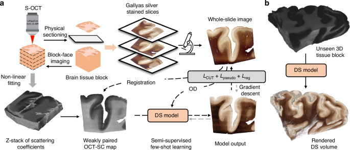
A major challenge in neuroscience is visualizing the structure of the human brain at different scales. Traditional histology reveals micro- and meso-scale brain features but suffers from staining variability, tissue damage, and distortion, which impedes accurate 3D reconstructions. The emerging label-free serial sectioning optical coherence tomography (S-OCT) technique offers uniform 3D imaging capability across samples but has poor histological interpretability despite its sensitivity to cortical features. Here, we present a novel 3D imaging framework that combines S-OCT with a deep-learning digital staining (DS) model. This enhanced imaging modality integrates high-throughput 3D imaging, low sample variability and high interpretability, making it suitable for 3D histology studies. We develop a novel semi-supervised learning technique to facilitate DS model training on weakly paired images for translating S-OCT to Gallyas silver staining. We demonstrate DS on various human cerebral cortex samples, achieving consistent staining quality and enhancing contrast across cortical layer boundaries. Additionally, we show that DS preserves geometry in 3D on cubic-centimeter tissue blocks, allowing for visualization of meso-scale vessel networks in the white matter. We believe that our technique has the potential for high-throughput, multiscale imaging of brain tissues and may facilitate studies of brain structures.
 求助内容:
求助内容: 应助结果提醒方式:


