{"title":"肺长距离:COVID-19肺炎后肺组织的组织病理学改变。","authors":"Grgur Salai, Jasna Tekavec-Trkanjec, Ivona Kovačević, Čedna Tomasović-Lončarić, Arijana Pačić, Mirna Vergles, Đivo Ljubičić, Daria Cvetković-Kučić, Ivica Lukšić, Bruno Baršić","doi":"","DOIUrl":null,"url":null,"abstract":"<p><strong>Aim: </strong>To investigate histopathological changes in the lung tissue of long-COVID patients.</p><p><strong>Methods: </strong>In this cross-sectional study, transbronchial lung biopsy was performed in long-COVID patients with persisting symptoms and radiological abnormalities. Histopathologic analyses were performed by using hematoxylin-eosin, Martius, Scarlet and Blue, Movat's, thyroid transcription factor 1, CD34, and CD68 staining.</p><p><strong>Results: </strong>Adequate biopsy samples were obtained from 29/32 patients. The median (Q1-Q3) time from disease onset to biopsy was 13 (9-20) weeks. We observed several histopathologic patterns: DAD with vascular abnormalities (VA) (n=8); VA with inflammatory pattern (n=4); inflammatory pattern (n=13), and fibrotic pattern (n=4). VA included capillary thrombi, dilated venules, and dissection of small pulmonary arteries. DAD with VA was detected up to the 9th week from the onset of disease; inflammatory pattern from the 8th to 28th week (4 patients with this pattern biopsied in the 11th-13th week had accompanying VA); and a predominantly fibrotic pattern was found at weeks 8, 10, 48, and 49.</p><p><strong>Conclusion: </strong>Our study observed a slow recovery of lung tissue with long-lasting DAD and VA, likely followed by interstitial inflammation or focal fibrosis. These findings might be the underlying cause of the slow recovery of long-COVID patients.</p>","PeriodicalId":10796,"journal":{"name":"Croatian Medical Journal","volume":"65 6","pages":"501-509"},"PeriodicalIF":2.3000,"publicationDate":"2024-12-30","publicationTypes":"Journal Article","fieldsOfStudy":null,"isOpenAccess":false,"openAccessPdf":"https://www.ncbi.nlm.nih.gov/pmc/articles/PMC11748447/pdf/","citationCount":"0","resultStr":"{\"title\":\"Lung long distance: histopathological changes in lung tissue after COVID-19 pneumonia.\",\"authors\":\"Grgur Salai, Jasna Tekavec-Trkanjec, Ivona Kovačević, Čedna Tomasović-Lončarić, Arijana Pačić, Mirna Vergles, Đivo Ljubičić, Daria Cvetković-Kučić, Ivica Lukšić, Bruno Baršić\",\"doi\":\"\",\"DOIUrl\":null,\"url\":null,\"abstract\":\"<p><strong>Aim: </strong>To investigate histopathological changes in the lung tissue of long-COVID patients.</p><p><strong>Methods: </strong>In this cross-sectional study, transbronchial lung biopsy was performed in long-COVID patients with persisting symptoms and radiological abnormalities. Histopathologic analyses were performed by using hematoxylin-eosin, Martius, Scarlet and Blue, Movat's, thyroid transcription factor 1, CD34, and CD68 staining.</p><p><strong>Results: </strong>Adequate biopsy samples were obtained from 29/32 patients. The median (Q1-Q3) time from disease onset to biopsy was 13 (9-20) weeks. We observed several histopathologic patterns: DAD with vascular abnormalities (VA) (n=8); VA with inflammatory pattern (n=4); inflammatory pattern (n=13), and fibrotic pattern (n=4). VA included capillary thrombi, dilated venules, and dissection of small pulmonary arteries. DAD with VA was detected up to the 9th week from the onset of disease; inflammatory pattern from the 8th to 28th week (4 patients with this pattern biopsied in the 11th-13th week had accompanying VA); and a predominantly fibrotic pattern was found at weeks 8, 10, 48, and 49.</p><p><strong>Conclusion: </strong>Our study observed a slow recovery of lung tissue with long-lasting DAD and VA, likely followed by interstitial inflammation or focal fibrosis. These findings might be the underlying cause of the slow recovery of long-COVID patients.</p>\",\"PeriodicalId\":10796,\"journal\":{\"name\":\"Croatian Medical Journal\",\"volume\":\"65 6\",\"pages\":\"501-509\"},\"PeriodicalIF\":2.3000,\"publicationDate\":\"2024-12-30\",\"publicationTypes\":\"Journal Article\",\"fieldsOfStudy\":null,\"isOpenAccess\":false,\"openAccessPdf\":\"https://www.ncbi.nlm.nih.gov/pmc/articles/PMC11748447/pdf/\",\"citationCount\":\"0\",\"resultStr\":null,\"platform\":\"Semanticscholar\",\"paperid\":null,\"PeriodicalName\":\"Croatian Medical Journal\",\"FirstCategoryId\":\"3\",\"ListUrlMain\":\"\",\"RegionNum\":4,\"RegionCategory\":\"医学\",\"ArticlePicture\":[],\"TitleCN\":null,\"AbstractTextCN\":null,\"PMCID\":null,\"EPubDate\":\"\",\"PubModel\":\"\",\"JCR\":\"Q2\",\"JCRName\":\"MEDICINE, GENERAL & INTERNAL\",\"Score\":null,\"Total\":0}","platform":"Semanticscholar","paperid":null,"PeriodicalName":"Croatian Medical Journal","FirstCategoryId":"3","ListUrlMain":"","RegionNum":4,"RegionCategory":"医学","ArticlePicture":[],"TitleCN":null,"AbstractTextCN":null,"PMCID":null,"EPubDate":"","PubModel":"","JCR":"Q2","JCRName":"MEDICINE, GENERAL & INTERNAL","Score":null,"Total":0}
引用次数: 0
摘要
目的:探讨长期新冠肺炎患者肺组织的病理变化。方法:在本横断面研究中,对持续症状和影像学异常的长期covid患者进行经支气管肺活检。采用苏木精-伊红、Martius、Scarlet and Blue、Movat’s、甲状腺转录因子1、CD34和CD68染色进行组织病理学分析。结果:32例患者中有29例获得了足够的活检样本。从发病到活检的中位时间(Q1-Q3)为13(9-20)周。我们观察到几种组织病理学模式:DAD伴血管异常(VA) (n=8);伴有炎症的VA (n=4);炎症型(n=13)和纤维化型(n=4)。VA包括毛细血管血栓、小静脉扩张和小肺动脉剥离。DAD合并VA在发病后第9周检测到;第8 ~ 28周的炎症模式(11 ~ 13周活检的4例伴VA);在第8周、第10周、第48周和第49周发现了主要的纤维化模式。结论:我们的研究观察到长期DAD和VA的肺组织恢复缓慢,可能随后出现间质炎症或局灶性纤维化。这些发现可能是长期患者恢复缓慢的根本原因。
Lung long distance: histopathological changes in lung tissue after COVID-19 pneumonia.
Aim: To investigate histopathological changes in the lung tissue of long-COVID patients.
Methods: In this cross-sectional study, transbronchial lung biopsy was performed in long-COVID patients with persisting symptoms and radiological abnormalities. Histopathologic analyses were performed by using hematoxylin-eosin, Martius, Scarlet and Blue, Movat's, thyroid transcription factor 1, CD34, and CD68 staining.
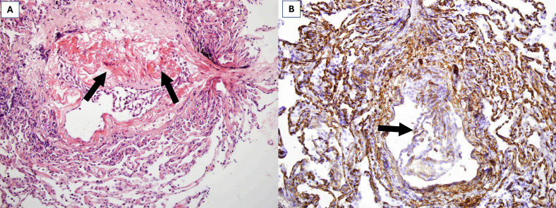
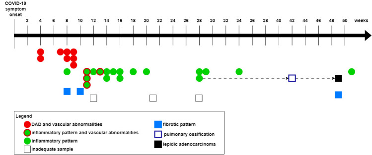
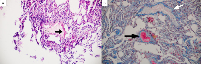
Results: Adequate biopsy samples were obtained from 29/32 patients. The median (Q1-Q3) time from disease onset to biopsy was 13 (9-20) weeks. We observed several histopathologic patterns: DAD with vascular abnormalities (VA) (n=8); VA with inflammatory pattern (n=4); inflammatory pattern (n=13), and fibrotic pattern (n=4). VA included capillary thrombi, dilated venules, and dissection of small pulmonary arteries. DAD with VA was detected up to the 9th week from the onset of disease; inflammatory pattern from the 8th to 28th week (4 patients with this pattern biopsied in the 11th-13th week had accompanying VA); and a predominantly fibrotic pattern was found at weeks 8, 10, 48, and 49.
Conclusion: Our study observed a slow recovery of lung tissue with long-lasting DAD and VA, likely followed by interstitial inflammation or focal fibrosis. These findings might be the underlying cause of the slow recovery of long-COVID patients.
期刊介绍:
Croatian Medical Journal (CMJ) is an international peer reviewed journal open to scientists from all fields of biomedicine and health related research.
Although CMJ welcomes all contributions that increase and expand on medical knowledge, the two areas are of the special interest: topics globally relevant for biomedicine and health and medicine in developing and emerging countries.

 求助内容:
求助内容: 应助结果提醒方式:


