{"title":"青蒿素通过激活5′- amp活化蛋白激酶(AMPK)保护人支气管上皮细胞免受胺碘酮诱导的氧化损伤。","authors":"Chao Yang, Wenjun Xiong, Jiayi Dong, Xia Zhao, Guang Liang, Wenhua Zheng","doi":"10.1080/13510002.2024.2447721","DOIUrl":null,"url":null,"abstract":"<p><strong>Background: </strong>Amiodarone, a common antiarrhythmic drug, is known for its severe side effects, including pulmonary toxicity, which involves oxidative stress and apoptosis. Artemisinin, an antimalarial drug, has shown cytoprotective properties by inhibiting oxidative stress and apoptosis. This study investigated the protective effects of artemisinin against amiodarone-induced toxicity in human bronchial epithelial cells (BEAS-2B) and mouse models.</p><p><strong>Results: </strong><i>In vitro</i> experiments revealed that amiodarone decreased cell viability, increased LDH release, ROS generation, caspase 3 activation, and apoptosis in BEAS-2B cells. Artemisinin counteracted these effects by upregulating p-AMPK, CaMKK2, Nrf2, and SOD1 protein levels, thereby protecting the cells from oxidative damage. The protective effect of artemisinin was diminished by the AMPK inhibitor Compound C or AMPKα knockdown. <i>In vivo</i> experiments demonstrated that artemisinin increased p-AMPK and Nrf2 protein levels in lung tissues, protecting against amiodarone-induced apoptosis and bronchial epithelial cell shedding in mice.</p><p><strong>Conclusion: </strong>These findings suggest that artemisinin protects airway epithelial cells and lung tissue from amiodarone-induced oxidative stress and apoptosis through AMPK activation, offering potential new strategies for preventing and treating amiodarone-induced pulmonary toxicity.</p>","PeriodicalId":21096,"journal":{"name":"Redox Report","volume":"30 1","pages":"2447721"},"PeriodicalIF":7.4000,"publicationDate":"2025-12-01","publicationTypes":"Journal Article","fieldsOfStudy":null,"isOpenAccess":false,"openAccessPdf":"https://www.ncbi.nlm.nih.gov/pmc/articles/PMC11731350/pdf/","citationCount":"0","resultStr":"{\"title\":\"Artemisinin protected human bronchial epithelial cells from amiodarone-induced oxidative damage via 5'-AMP-activated protein kinase (AMPK) activation.\",\"authors\":\"Chao Yang, Wenjun Xiong, Jiayi Dong, Xia Zhao, Guang Liang, Wenhua Zheng\",\"doi\":\"10.1080/13510002.2024.2447721\",\"DOIUrl\":null,\"url\":null,\"abstract\":\"<p><strong>Background: </strong>Amiodarone, a common antiarrhythmic drug, is known for its severe side effects, including pulmonary toxicity, which involves oxidative stress and apoptosis. Artemisinin, an antimalarial drug, has shown cytoprotective properties by inhibiting oxidative stress and apoptosis. This study investigated the protective effects of artemisinin against amiodarone-induced toxicity in human bronchial epithelial cells (BEAS-2B) and mouse models.</p><p><strong>Results: </strong><i>In vitro</i> experiments revealed that amiodarone decreased cell viability, increased LDH release, ROS generation, caspase 3 activation, and apoptosis in BEAS-2B cells. Artemisinin counteracted these effects by upregulating p-AMPK, CaMKK2, Nrf2, and SOD1 protein levels, thereby protecting the cells from oxidative damage. The protective effect of artemisinin was diminished by the AMPK inhibitor Compound C or AMPKα knockdown. <i>In vivo</i> experiments demonstrated that artemisinin increased p-AMPK and Nrf2 protein levels in lung tissues, protecting against amiodarone-induced apoptosis and bronchial epithelial cell shedding in mice.</p><p><strong>Conclusion: </strong>These findings suggest that artemisinin protects airway epithelial cells and lung tissue from amiodarone-induced oxidative stress and apoptosis through AMPK activation, offering potential new strategies for preventing and treating amiodarone-induced pulmonary toxicity.</p>\",\"PeriodicalId\":21096,\"journal\":{\"name\":\"Redox Report\",\"volume\":\"30 1\",\"pages\":\"2447721\"},\"PeriodicalIF\":7.4000,\"publicationDate\":\"2025-12-01\",\"publicationTypes\":\"Journal Article\",\"fieldsOfStudy\":null,\"isOpenAccess\":false,\"openAccessPdf\":\"https://www.ncbi.nlm.nih.gov/pmc/articles/PMC11731350/pdf/\",\"citationCount\":\"0\",\"resultStr\":null,\"platform\":\"Semanticscholar\",\"paperid\":null,\"PeriodicalName\":\"Redox Report\",\"FirstCategoryId\":\"99\",\"ListUrlMain\":\"https://doi.org/10.1080/13510002.2024.2447721\",\"RegionNum\":2,\"RegionCategory\":\"生物学\",\"ArticlePicture\":[],\"TitleCN\":null,\"AbstractTextCN\":null,\"PMCID\":null,\"EPubDate\":\"2025/1/13 0:00:00\",\"PubModel\":\"Epub\",\"JCR\":\"Q1\",\"JCRName\":\"BIOCHEMISTRY & MOLECULAR BIOLOGY\",\"Score\":null,\"Total\":0}","platform":"Semanticscholar","paperid":null,"PeriodicalName":"Redox Report","FirstCategoryId":"99","ListUrlMain":"https://doi.org/10.1080/13510002.2024.2447721","RegionNum":2,"RegionCategory":"生物学","ArticlePicture":[],"TitleCN":null,"AbstractTextCN":null,"PMCID":null,"EPubDate":"2025/1/13 0:00:00","PubModel":"Epub","JCR":"Q1","JCRName":"BIOCHEMISTRY & MOLECULAR BIOLOGY","Score":null,"Total":0}
Artemisinin protected human bronchial epithelial cells from amiodarone-induced oxidative damage via 5'-AMP-activated protein kinase (AMPK) activation.
Background: Amiodarone, a common antiarrhythmic drug, is known for its severe side effects, including pulmonary toxicity, which involves oxidative stress and apoptosis. Artemisinin, an antimalarial drug, has shown cytoprotective properties by inhibiting oxidative stress and apoptosis. This study investigated the protective effects of artemisinin against amiodarone-induced toxicity in human bronchial epithelial cells (BEAS-2B) and mouse models.
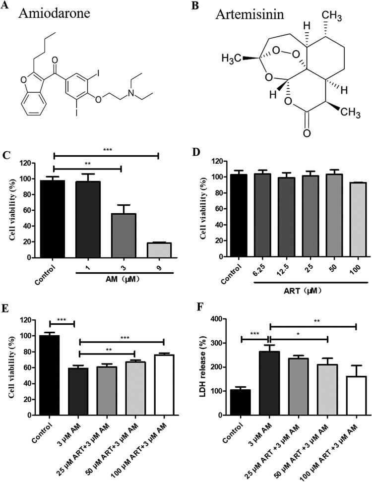
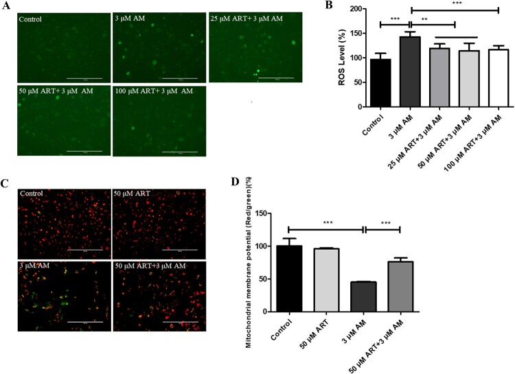
Results: In vitro experiments revealed that amiodarone decreased cell viability, increased LDH release, ROS generation, caspase 3 activation, and apoptosis in BEAS-2B cells. Artemisinin counteracted these effects by upregulating p-AMPK, CaMKK2, Nrf2, and SOD1 protein levels, thereby protecting the cells from oxidative damage. The protective effect of artemisinin was diminished by the AMPK inhibitor Compound C or AMPKα knockdown. In vivo experiments demonstrated that artemisinin increased p-AMPK and Nrf2 protein levels in lung tissues, protecting against amiodarone-induced apoptosis and bronchial epithelial cell shedding in mice.
Conclusion: These findings suggest that artemisinin protects airway epithelial cells and lung tissue from amiodarone-induced oxidative stress and apoptosis through AMPK activation, offering potential new strategies for preventing and treating amiodarone-induced pulmonary toxicity.
期刊介绍:
Redox Report is a multidisciplinary peer-reviewed open access journal focusing on the role of free radicals, oxidative stress, activated oxygen, perioxidative and redox processes, primarily in the human environment and human pathology. Relevant papers on the animal and plant environment, biology and pathology will also be included.
While emphasis is placed upon methodological and intellectual advances underpinned by new data, the journal offers scope for review, hypotheses, critiques and other forms of discussion.
 求助内容:
求助内容: 应助结果提醒方式:


