Jill Halstead, Carmen Martín-Hervás, Elizabeth M A Hensor, Anne-Maree Keenan, Philip G Conaghan, Dennis McGonagle, John B Arnold, Jennifer Jones, Anthony C Redmond
{"title":"中足痛患者的临床和mri检测成像结果之间的关系,一项横断面研究。","authors":"Jill Halstead, Carmen Martín-Hervás, Elizabeth M A Hensor, Anne-Maree Keenan, Philip G Conaghan, Dennis McGonagle, John B Arnold, Jennifer Jones, Anthony C Redmond","doi":"10.1002/jfa2.70019","DOIUrl":null,"url":null,"abstract":"<p><strong>Background: </strong>Midfoot pain is common but poorly understood, with radiographs often indicating no anomalies. This study aimed to describe bone, joint and soft tissue changes and to explore associations between MRI-detected abnormalities and clinical symptoms (pain and disability) in a group of adults with midfoot pain, but who were radiographically negative for osteoarthritis.</p><p><strong>Methods: </strong>Community-based participants with midfoot pain underwent an MRI scan of one foot and scored semi-quantitatively using the Foot OsteoArthritis MRI Score (FOAMRIS). Foot pain and disability were recorded using visual analog scales (VAS) and the Modified-Manchester Foot Pain Disability Index (MMFPDI). Associations were assessed for continuous data using Spearman's Rho, and for categorical data, a Wilcoxon signed rank test. Linear regression was used to explore the association between participant-reported measures and MRI abnormalities, adjusted for age, sex and BMI.</p><p><strong>Results: </strong>Sixty-one participants (70% female, mean age 48.5 years, median BMI 28.6 kg/m<sup>2</sup>) were included. Median VAS pain was 31/100 mm (IQR 21-47) and median disability was 30/48 (IQR 26-36). There was a moderate association between midfoot pain severity and the number of joints exhibiting joint space narrowing; adjusted results suggested 31% (95% confidence interval 3%-68%) worse VAS pain with each additional affected joint. Greater numbers of joints with cysts were associated with worse VAS pain [14% (0%-31%)] and disability [1.1 units (0-2.2)]. Effusion/synovitis was associated with MMFPDI pain. No other MRI abnormalities were associated with sex, body mass and foot pain/disability measures. Bone marrow lesions, joint space narrowing, cysts and osteophytes occurred more frequently with age. MRI abnormalities were common, particularly in the talo-navicular joint, first and second cuneo-metatarsal joints. Those with dorsal foot pain had more multi-joint involvement, bone marrow lesions, joint space narrowing and cysts and for those with pain on midfoot movement, bone marrow lesions and cysts were reported.</p><p><strong>Conclusions: </strong>In people with midfoot pain, MRI-detected features of osteoarthritis and soft-tissue abnormalities were found, clustered in the medial and intermediate cuneiform joints. These features were more common with age but not associated with pain or disability measures. Younger people with dorsal midfoot pain exhibited early signs of bone and joint features of osteoarthritis and we recommend further imaging studies to determine the clinical and diagnostic significance.</p>","PeriodicalId":49164,"journal":{"name":"Journal of Foot and Ankle Research","volume":"18 1","pages":"e70019"},"PeriodicalIF":2.2000,"publicationDate":"2025-03-01","publicationTypes":"Journal Article","fieldsOfStudy":null,"isOpenAccess":false,"openAccessPdf":"https://www.ncbi.nlm.nih.gov/pmc/articles/PMC11724207/pdf/","citationCount":"0","resultStr":"{\"title\":\"Association between clinical and MRI-detected imaging findings for people with midfoot pain, a cross-sectional study.\",\"authors\":\"Jill Halstead, Carmen Martín-Hervás, Elizabeth M A Hensor, Anne-Maree Keenan, Philip G Conaghan, Dennis McGonagle, John B Arnold, Jennifer Jones, Anthony C Redmond\",\"doi\":\"10.1002/jfa2.70019\",\"DOIUrl\":null,\"url\":null,\"abstract\":\"<p><strong>Background: </strong>Midfoot pain is common but poorly understood, with radiographs often indicating no anomalies. This study aimed to describe bone, joint and soft tissue changes and to explore associations between MRI-detected abnormalities and clinical symptoms (pain and disability) in a group of adults with midfoot pain, but who were radiographically negative for osteoarthritis.</p><p><strong>Methods: </strong>Community-based participants with midfoot pain underwent an MRI scan of one foot and scored semi-quantitatively using the Foot OsteoArthritis MRI Score (FOAMRIS). Foot pain and disability were recorded using visual analog scales (VAS) and the Modified-Manchester Foot Pain Disability Index (MMFPDI). Associations were assessed for continuous data using Spearman's Rho, and for categorical data, a Wilcoxon signed rank test. Linear regression was used to explore the association between participant-reported measures and MRI abnormalities, adjusted for age, sex and BMI.</p><p><strong>Results: </strong>Sixty-one participants (70% female, mean age 48.5 years, median BMI 28.6 kg/m<sup>2</sup>) were included. Median VAS pain was 31/100 mm (IQR 21-47) and median disability was 30/48 (IQR 26-36). There was a moderate association between midfoot pain severity and the number of joints exhibiting joint space narrowing; adjusted results suggested 31% (95% confidence interval 3%-68%) worse VAS pain with each additional affected joint. Greater numbers of joints with cysts were associated with worse VAS pain [14% (0%-31%)] and disability [1.1 units (0-2.2)]. Effusion/synovitis was associated with MMFPDI pain. No other MRI abnormalities were associated with sex, body mass and foot pain/disability measures. Bone marrow lesions, joint space narrowing, cysts and osteophytes occurred more frequently with age. MRI abnormalities were common, particularly in the talo-navicular joint, first and second cuneo-metatarsal joints. Those with dorsal foot pain had more multi-joint involvement, bone marrow lesions, joint space narrowing and cysts and for those with pain on midfoot movement, bone marrow lesions and cysts were reported.</p><p><strong>Conclusions: </strong>In people with midfoot pain, MRI-detected features of osteoarthritis and soft-tissue abnormalities were found, clustered in the medial and intermediate cuneiform joints. These features were more common with age but not associated with pain or disability measures. Younger people with dorsal midfoot pain exhibited early signs of bone and joint features of osteoarthritis and we recommend further imaging studies to determine the clinical and diagnostic significance.</p>\",\"PeriodicalId\":49164,\"journal\":{\"name\":\"Journal of Foot and Ankle Research\",\"volume\":\"18 1\",\"pages\":\"e70019\"},\"PeriodicalIF\":2.2000,\"publicationDate\":\"2025-03-01\",\"publicationTypes\":\"Journal Article\",\"fieldsOfStudy\":null,\"isOpenAccess\":false,\"openAccessPdf\":\"https://www.ncbi.nlm.nih.gov/pmc/articles/PMC11724207/pdf/\",\"citationCount\":\"0\",\"resultStr\":null,\"platform\":\"Semanticscholar\",\"paperid\":null,\"PeriodicalName\":\"Journal of Foot and Ankle Research\",\"FirstCategoryId\":\"3\",\"ListUrlMain\":\"https://doi.org/10.1002/jfa2.70019\",\"RegionNum\":3,\"RegionCategory\":\"医学\",\"ArticlePicture\":[],\"TitleCN\":null,\"AbstractTextCN\":null,\"PMCID\":null,\"EPubDate\":\"\",\"PubModel\":\"\",\"JCR\":\"Q1\",\"JCRName\":\"ORTHOPEDICS\",\"Score\":null,\"Total\":0}","platform":"Semanticscholar","paperid":null,"PeriodicalName":"Journal of Foot and Ankle Research","FirstCategoryId":"3","ListUrlMain":"https://doi.org/10.1002/jfa2.70019","RegionNum":3,"RegionCategory":"医学","ArticlePicture":[],"TitleCN":null,"AbstractTextCN":null,"PMCID":null,"EPubDate":"","PubModel":"","JCR":"Q1","JCRName":"ORTHOPEDICS","Score":null,"Total":0}
引用次数: 0
摘要
背景:足中部疼痛是一种常见的疼痛,但人们对其了解甚少,x线片通常显示无异常。本研究旨在描述骨、关节和软组织的变化,并探讨mri检测到的异常与临床症状(疼痛和残疾)之间的关系,这些患者患有足中部疼痛,但影像学检查为骨关节炎阴性。方法:以社区为基础的中足疼痛参与者对一只脚进行MRI扫描,并使用足骨关节炎MRI评分(FOAMRIS)进行半定量评分。采用视觉模拟量表(VAS)和改良曼彻斯特足部疼痛残疾指数(MMFPDI)记录足部疼痛和残疾情况。使用Spearman’s Rho对连续数据进行关联评估,使用Wilcoxon符号秩检验对分类数据进行关联评估。线性回归用于探讨参与者报告的测量与MRI异常之间的关系,并根据年龄、性别和BMI进行调整。结果:纳入61名参与者(70%为女性,平均年龄48.5岁,中位BMI 28.6 kg/m2)。VAS疼痛中位数为31/100 mm (IQR 21-47),残疾中位数为30/48 (IQR 26-36)。足中部疼痛的严重程度与关节间隙变窄的关节数量有中度相关性;调整后的结果显示,每增加一个受影响的关节,VAS疼痛加重31%(95%置信区间为3%-68%)。有囊肿的关节越多,VAS疼痛就越严重[14%(0%-31%)],残疾也越严重[1.1个单位(0-2.2)]。积液/滑膜炎与MMFPDI疼痛相关。没有其他MRI异常与性别、体重和足部疼痛/残疾措施相关。随着年龄的增长,骨髓病变、关节间隙狭窄、囊肿和骨赘的发生频率增加。MRI异常是常见的,特别是在距舟关节,第一和第二楔跖关节。足背疼痛患者有更多的多关节受累、骨髓病变、关节间隙狭窄和囊肿,而足中运动疼痛患者则有骨髓病变和囊肿的报道。结论:在足中部疼痛患者中,mri检测到骨关节炎和软组织异常的特征,集中在内侧和中间楔形关节。这些特征在年龄上更常见,但与疼痛或残疾措施无关。年轻的足背中痛患者表现出骨关节炎的早期骨和关节特征,我们建议进一步的影像学检查以确定临床和诊断意义。
Association between clinical and MRI-detected imaging findings for people with midfoot pain, a cross-sectional study.
Background: Midfoot pain is common but poorly understood, with radiographs often indicating no anomalies. This study aimed to describe bone, joint and soft tissue changes and to explore associations between MRI-detected abnormalities and clinical symptoms (pain and disability) in a group of adults with midfoot pain, but who were radiographically negative for osteoarthritis.
Methods: Community-based participants with midfoot pain underwent an MRI scan of one foot and scored semi-quantitatively using the Foot OsteoArthritis MRI Score (FOAMRIS). Foot pain and disability were recorded using visual analog scales (VAS) and the Modified-Manchester Foot Pain Disability Index (MMFPDI). Associations were assessed for continuous data using Spearman's Rho, and for categorical data, a Wilcoxon signed rank test. Linear regression was used to explore the association between participant-reported measures and MRI abnormalities, adjusted for age, sex and BMI.
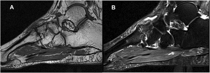
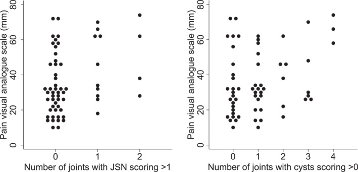
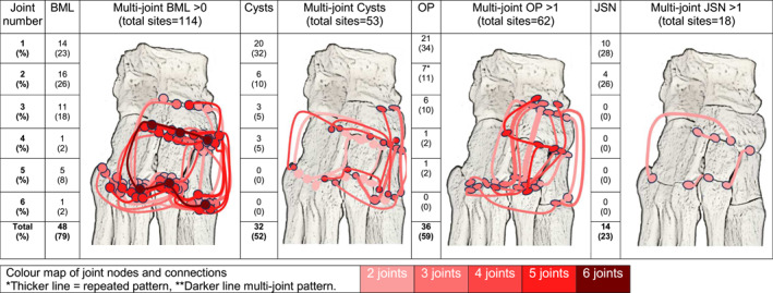
Results: Sixty-one participants (70% female, mean age 48.5 years, median BMI 28.6 kg/m2) were included. Median VAS pain was 31/100 mm (IQR 21-47) and median disability was 30/48 (IQR 26-36). There was a moderate association between midfoot pain severity and the number of joints exhibiting joint space narrowing; adjusted results suggested 31% (95% confidence interval 3%-68%) worse VAS pain with each additional affected joint. Greater numbers of joints with cysts were associated with worse VAS pain [14% (0%-31%)] and disability [1.1 units (0-2.2)]. Effusion/synovitis was associated with MMFPDI pain. No other MRI abnormalities were associated with sex, body mass and foot pain/disability measures. Bone marrow lesions, joint space narrowing, cysts and osteophytes occurred more frequently with age. MRI abnormalities were common, particularly in the talo-navicular joint, first and second cuneo-metatarsal joints. Those with dorsal foot pain had more multi-joint involvement, bone marrow lesions, joint space narrowing and cysts and for those with pain on midfoot movement, bone marrow lesions and cysts were reported.
Conclusions: In people with midfoot pain, MRI-detected features of osteoarthritis and soft-tissue abnormalities were found, clustered in the medial and intermediate cuneiform joints. These features were more common with age but not associated with pain or disability measures. Younger people with dorsal midfoot pain exhibited early signs of bone and joint features of osteoarthritis and we recommend further imaging studies to determine the clinical and diagnostic significance.
期刊介绍:
Journal of Foot and Ankle Research, the official journal of the Australian Podiatry Association and The College of Podiatry (UK), is an open access journal that encompasses all aspects of policy, organisation, delivery and clinical practice related to the assessment, diagnosis, prevention and management of foot and ankle disorders.
Journal of Foot and Ankle Research covers a wide range of clinical subject areas, including diabetology, paediatrics, sports medicine, gerontology and geriatrics, foot surgery, physical therapy, dermatology, wound management, radiology, biomechanics and bioengineering, orthotics and prosthetics, as well the broad areas of epidemiology, policy, organisation and delivery of services related to foot and ankle care.
The journal encourages submissions from all health professionals who manage lower limb conditions, including podiatrists, nurses, physical therapists and physiotherapists, orthopaedists, manual therapists, medical specialists and general medical practitioners, as well as health service researchers concerned with foot and ankle care.
The Australian Podiatry Association and the College of Podiatry (UK) have reserve funds to cover the article-processing charge for manuscripts submitted by its members. Society members can email the appropriate contact at Australian Podiatry Association or The College of Podiatry to obtain the corresponding code to enter on submission.
 求助内容:
求助内容: 应助结果提醒方式:


