Nurcan Özyurt Koçakoğlu, Doğan Erhan Ersoy, Hicret Arslan, Selami Candan
{"title":"金龟甲科金龟甲的中肠和马尔匹氏小管的解剖和组织学。","authors":"Nurcan Özyurt Koçakoğlu, Doğan Erhan Ersoy, Hicret Arslan, Selami Candan","doi":"10.1007/s00709-024-02021-1","DOIUrl":null,"url":null,"abstract":"<p><p>Copris are part of the Scarabaeidae family of Coleoptera. Copris are dung beetles or coprophagous beetles. These insects are called tunnelers because they excavate channels in the substrate. They use dead organisms and non-living organic compounds as a nutrient source. By breaking down dead matter, they provide nutrients that are important to the environment and necessary for the survival of other organisms. No studies have yet examined the midgut structure and Malpighian tubules of Copris. Therefore, this study investigated the histo-anatomical structure of the midgut and Malpighian tubules of Copris felschei Reitter, 1892 (Coleoptera: Scarabaeidae) using light and scanning electron microscopy (SEM) in detail. The midgut of C. felschei represents the largest part of the alimentary canal. Muscle layers and a monolayer of cylindrical epithelium surround the midgut wall. A peritrophic membrane envelops food in the midgut lumen, and crystals were observed within the lumen. The surface of the midgut has regenerative crypts and tracheae. The Malpighian tubules are arranged in two pairs and connect proximally between the midgut and hindgut. The Malpighian tubules are composed of a single layer of cuboidal epithelium. Numerous balloon-like tracheae were observed interspersed between the midgut and Malpighian tubules. Light and SEM images of the tracheae reveal a spongy structure with hollow chambers. These findings are anticipated to advance future research and deepen our understanding of the alimentary canal in Coleoptera, particularly within the Scarabaeidae family.</p>","PeriodicalId":20731,"journal":{"name":"Protoplasma","volume":" ","pages":"683-693"},"PeriodicalIF":2.5000,"publicationDate":"2025-05-01","publicationTypes":"Journal Article","fieldsOfStudy":null,"isOpenAccess":false,"openAccessPdf":"https://www.ncbi.nlm.nih.gov/pmc/articles/PMC12018526/pdf/","citationCount":"0","resultStr":"{\"title\":\"The anatomy and histology of the midgut and Malpighian tubules of Copris felschei Reitter, 1892 (Coleoptera: Scarabaeidae).\",\"authors\":\"Nurcan Özyurt Koçakoğlu, Doğan Erhan Ersoy, Hicret Arslan, Selami Candan\",\"doi\":\"10.1007/s00709-024-02021-1\",\"DOIUrl\":null,\"url\":null,\"abstract\":\"<p><p>Copris are part of the Scarabaeidae family of Coleoptera. Copris are dung beetles or coprophagous beetles. These insects are called tunnelers because they excavate channels in the substrate. They use dead organisms and non-living organic compounds as a nutrient source. By breaking down dead matter, they provide nutrients that are important to the environment and necessary for the survival of other organisms. No studies have yet examined the midgut structure and Malpighian tubules of Copris. Therefore, this study investigated the histo-anatomical structure of the midgut and Malpighian tubules of Copris felschei Reitter, 1892 (Coleoptera: Scarabaeidae) using light and scanning electron microscopy (SEM) in detail. The midgut of C. felschei represents the largest part of the alimentary canal. Muscle layers and a monolayer of cylindrical epithelium surround the midgut wall. A peritrophic membrane envelops food in the midgut lumen, and crystals were observed within the lumen. The surface of the midgut has regenerative crypts and tracheae. The Malpighian tubules are arranged in two pairs and connect proximally between the midgut and hindgut. The Malpighian tubules are composed of a single layer of cuboidal epithelium. Numerous balloon-like tracheae were observed interspersed between the midgut and Malpighian tubules. Light and SEM images of the tracheae reveal a spongy structure with hollow chambers. These findings are anticipated to advance future research and deepen our understanding of the alimentary canal in Coleoptera, particularly within the Scarabaeidae family.</p>\",\"PeriodicalId\":20731,\"journal\":{\"name\":\"Protoplasma\",\"volume\":\" \",\"pages\":\"683-693\"},\"PeriodicalIF\":2.5000,\"publicationDate\":\"2025-05-01\",\"publicationTypes\":\"Journal Article\",\"fieldsOfStudy\":null,\"isOpenAccess\":false,\"openAccessPdf\":\"https://www.ncbi.nlm.nih.gov/pmc/articles/PMC12018526/pdf/\",\"citationCount\":\"0\",\"resultStr\":null,\"platform\":\"Semanticscholar\",\"paperid\":null,\"PeriodicalName\":\"Protoplasma\",\"FirstCategoryId\":\"99\",\"ListUrlMain\":\"https://doi.org/10.1007/s00709-024-02021-1\",\"RegionNum\":3,\"RegionCategory\":\"生物学\",\"ArticlePicture\":[],\"TitleCN\":null,\"AbstractTextCN\":null,\"PMCID\":null,\"EPubDate\":\"2025/1/11 0:00:00\",\"PubModel\":\"Epub\",\"JCR\":\"Q3\",\"JCRName\":\"CELL BIOLOGY\",\"Score\":null,\"Total\":0}","platform":"Semanticscholar","paperid":null,"PeriodicalName":"Protoplasma","FirstCategoryId":"99","ListUrlMain":"https://doi.org/10.1007/s00709-024-02021-1","RegionNum":3,"RegionCategory":"生物学","ArticlePicture":[],"TitleCN":null,"AbstractTextCN":null,"PMCID":null,"EPubDate":"2025/1/11 0:00:00","PubModel":"Epub","JCR":"Q3","JCRName":"CELL BIOLOGY","Score":null,"Total":0}
The anatomy and histology of the midgut and Malpighian tubules of Copris felschei Reitter, 1892 (Coleoptera: Scarabaeidae).
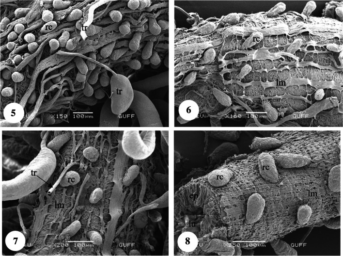
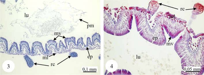
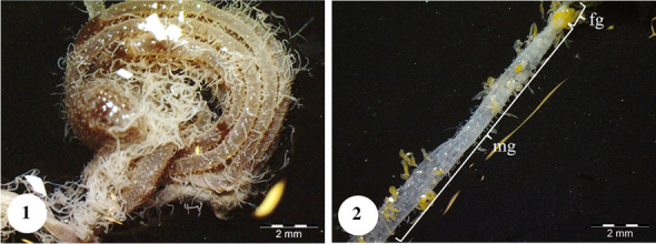
Copris are part of the Scarabaeidae family of Coleoptera. Copris are dung beetles or coprophagous beetles. These insects are called tunnelers because they excavate channels in the substrate. They use dead organisms and non-living organic compounds as a nutrient source. By breaking down dead matter, they provide nutrients that are important to the environment and necessary for the survival of other organisms. No studies have yet examined the midgut structure and Malpighian tubules of Copris. Therefore, this study investigated the histo-anatomical structure of the midgut and Malpighian tubules of Copris felschei Reitter, 1892 (Coleoptera: Scarabaeidae) using light and scanning electron microscopy (SEM) in detail. The midgut of C. felschei represents the largest part of the alimentary canal. Muscle layers and a monolayer of cylindrical epithelium surround the midgut wall. A peritrophic membrane envelops food in the midgut lumen, and crystals were observed within the lumen. The surface of the midgut has regenerative crypts and tracheae. The Malpighian tubules are arranged in two pairs and connect proximally between the midgut and hindgut. The Malpighian tubules are composed of a single layer of cuboidal epithelium. Numerous balloon-like tracheae were observed interspersed between the midgut and Malpighian tubules. Light and SEM images of the tracheae reveal a spongy structure with hollow chambers. These findings are anticipated to advance future research and deepen our understanding of the alimentary canal in Coleoptera, particularly within the Scarabaeidae family.
期刊介绍:
Protoplasma publishes original papers, short communications and review articles which are of interest to cell biology in all its scientific and applied aspects. We seek contributions dealing with plants and animals but also prokaryotes, protists and fungi, from the following fields:
cell biology of both single and multicellular organisms
molecular cytology
the cell cycle
membrane biology including biogenesis, dynamics, energetics and electrophysiology
inter- and intracellular transport
the cytoskeleton
organelles
experimental and quantitative ultrastructure
cyto- and histochemistry
Further, conceptual contributions such as new models or discoveries at the cutting edge of cell biology research will be published under the headings "New Ideas in Cell Biology".

 求助内容:
求助内容: 应助结果提醒方式:


