Rustamzhon Melikov, Giuseppina Iachetta, Marta d'Amora, Giovanni Melle, Silvia Conti, Francesco Tantussi, Michele Dipalo, Francesco De Angelis
{"title":"平面MEA电极对大鼠初级神经元自发电生理活动的纵向和无创细胞内记录","authors":"Rustamzhon Melikov, Giuseppina Iachetta, Marta d'Amora, Giovanni Melle, Silvia Conti, Francesco Tantussi, Michele Dipalo, Francesco De Angelis","doi":"10.1002/adma.202412697","DOIUrl":null,"url":null,"abstract":"<p>Presently, the in vitro recording of intracellular neuronal signals on microelectrode arrays (MEAs) requires complex 3D nanostructures or invasive and approaches such as electroporation. Here, it is shown that laser poration enables intracellular coupling on planar electrodes without damaging neurons or altering their spontaneous electrophysiological activity, allowing the process to be repeated multiple times on the same cells. This capability distinguishes laser-based neuron poration from more invasive methods like electroporation, which typically serve as endpoint measurement for cells. It is demonstrated that planar MEA electrodes, when combined with laser cell optoporation and live cell staining, can record spontaneous intracellular signaling from primary neurons in vitro. This approach allows for the detection of attenuated signals resembling positive monophasic intracellular action potentials. Recordings after laser optoporation also reveal subthreshold signals such as post-synaptic potentials that are essential for assessing neuronal network plasticity and connectivity. Moreover, the noninvasiveness of the process enables repeated intracellular recordings over multiple days from the same cells.</p>","PeriodicalId":114,"journal":{"name":"Advanced Materials","volume":"37 8","pages":""},"PeriodicalIF":26.8000,"publicationDate":"2025-01-10","publicationTypes":"Journal Article","fieldsOfStudy":null,"isOpenAccess":false,"openAccessPdf":"https://onlinelibrary.wiley.com/doi/epdf/10.1002/adma.202412697","citationCount":"0","resultStr":"{\"title\":\"Longitudinal and Noninvasive Intracellular Recordings of Spontaneous Electrophysiological Activity in Rat Primary Neurons on Planar MEA Electrodes\",\"authors\":\"Rustamzhon Melikov, Giuseppina Iachetta, Marta d'Amora, Giovanni Melle, Silvia Conti, Francesco Tantussi, Michele Dipalo, Francesco De Angelis\",\"doi\":\"10.1002/adma.202412697\",\"DOIUrl\":null,\"url\":null,\"abstract\":\"<p>Presently, the in vitro recording of intracellular neuronal signals on microelectrode arrays (MEAs) requires complex 3D nanostructures or invasive and approaches such as electroporation. Here, it is shown that laser poration enables intracellular coupling on planar electrodes without damaging neurons or altering their spontaneous electrophysiological activity, allowing the process to be repeated multiple times on the same cells. This capability distinguishes laser-based neuron poration from more invasive methods like electroporation, which typically serve as endpoint measurement for cells. It is demonstrated that planar MEA electrodes, when combined with laser cell optoporation and live cell staining, can record spontaneous intracellular signaling from primary neurons in vitro. This approach allows for the detection of attenuated signals resembling positive monophasic intracellular action potentials. Recordings after laser optoporation also reveal subthreshold signals such as post-synaptic potentials that are essential for assessing neuronal network plasticity and connectivity. Moreover, the noninvasiveness of the process enables repeated intracellular recordings over multiple days from the same cells.</p>\",\"PeriodicalId\":114,\"journal\":{\"name\":\"Advanced Materials\",\"volume\":\"37 8\",\"pages\":\"\"},\"PeriodicalIF\":26.8000,\"publicationDate\":\"2025-01-10\",\"publicationTypes\":\"Journal Article\",\"fieldsOfStudy\":null,\"isOpenAccess\":false,\"openAccessPdf\":\"https://onlinelibrary.wiley.com/doi/epdf/10.1002/adma.202412697\",\"citationCount\":\"0\",\"resultStr\":null,\"platform\":\"Semanticscholar\",\"paperid\":null,\"PeriodicalName\":\"Advanced Materials\",\"FirstCategoryId\":\"88\",\"ListUrlMain\":\"https://onlinelibrary.wiley.com/doi/10.1002/adma.202412697\",\"RegionNum\":1,\"RegionCategory\":\"材料科学\",\"ArticlePicture\":[],\"TitleCN\":null,\"AbstractTextCN\":null,\"PMCID\":null,\"EPubDate\":\"\",\"PubModel\":\"\",\"JCR\":\"Q1\",\"JCRName\":\"CHEMISTRY, MULTIDISCIPLINARY\",\"Score\":null,\"Total\":0}","platform":"Semanticscholar","paperid":null,"PeriodicalName":"Advanced Materials","FirstCategoryId":"88","ListUrlMain":"https://onlinelibrary.wiley.com/doi/10.1002/adma.202412697","RegionNum":1,"RegionCategory":"材料科学","ArticlePicture":[],"TitleCN":null,"AbstractTextCN":null,"PMCID":null,"EPubDate":"","PubModel":"","JCR":"Q1","JCRName":"CHEMISTRY, MULTIDISCIPLINARY","Score":null,"Total":0}
Longitudinal and Noninvasive Intracellular Recordings of Spontaneous Electrophysiological Activity in Rat Primary Neurons on Planar MEA Electrodes
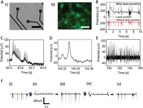
Presently, the in vitro recording of intracellular neuronal signals on microelectrode arrays (MEAs) requires complex 3D nanostructures or invasive and approaches such as electroporation. Here, it is shown that laser poration enables intracellular coupling on planar electrodes without damaging neurons or altering their spontaneous electrophysiological activity, allowing the process to be repeated multiple times on the same cells. This capability distinguishes laser-based neuron poration from more invasive methods like electroporation, which typically serve as endpoint measurement for cells. It is demonstrated that planar MEA electrodes, when combined with laser cell optoporation and live cell staining, can record spontaneous intracellular signaling from primary neurons in vitro. This approach allows for the detection of attenuated signals resembling positive monophasic intracellular action potentials. Recordings after laser optoporation also reveal subthreshold signals such as post-synaptic potentials that are essential for assessing neuronal network plasticity and connectivity. Moreover, the noninvasiveness of the process enables repeated intracellular recordings over multiple days from the same cells.
期刊介绍:
Advanced Materials, one of the world's most prestigious journals and the foundation of the Advanced portfolio, is the home of choice for best-in-class materials science for more than 30 years. Following this fast-growing and interdisciplinary field, we are considering and publishing the most important discoveries on any and all materials from materials scientists, chemists, physicists, engineers as well as health and life scientists and bringing you the latest results and trends in modern materials-related research every week.
 求助内容:
求助内容: 应助结果提醒方式:


