Baolong Zhang, Haiyan Yu, Dmytro Pylypenko, Jining Sun
{"title":"顶骨的结缔组织增生纤维瘤:病例报告及文献复习。","authors":"Baolong Zhang, Haiyan Yu, Dmytro Pylypenko, Jining Sun","doi":"10.25259/JCIS_136_2024","DOIUrl":null,"url":null,"abstract":"<p><p>Desmoplastic fibroma (DF) is an uncommon benign bone tumor that typically affects the facial bones, with cerebral cranium involvement being extremely rare. We report a unique case of DF in the parietal bone of a 28-year-old woman, notable for its rapid growth during pregnancy-a phenomenon not previously documented. The imaging features of this case also differ from all but one previously reported case. The patient underwent surgical removal, and histopathology confirmed the diagnosis of DF (collagenous fibroma). After 17 months of follow-up, no local recurrence was observed. We also provide a comprehensive review of 32 cases involving DF of the cerebral cranium, analyzing clinical features, imaging findings, treatment methods, and recurrence patterns. This case highlights the importance of considering DF in the differential diagnosis of cranial lesions, particularly in pregnant patients with rapid tumor growth. Complete surgical resection with a wide margin remains the recommended treatment to minimize recurrence risk.</p>","PeriodicalId":15512,"journal":{"name":"Journal of Clinical Imaging Science","volume":"14 ","pages":"48"},"PeriodicalIF":1.3000,"publicationDate":"2024-12-10","publicationTypes":"Journal Article","fieldsOfStudy":null,"isOpenAccess":false,"openAccessPdf":"https://www.ncbi.nlm.nih.gov/pmc/articles/PMC11704291/pdf/","citationCount":"0","resultStr":"{\"title\":\"Desmoplastic (collagenous) fibroma of the parietal bone: Case report and review of the literature.\",\"authors\":\"Baolong Zhang, Haiyan Yu, Dmytro Pylypenko, Jining Sun\",\"doi\":\"10.25259/JCIS_136_2024\",\"DOIUrl\":null,\"url\":null,\"abstract\":\"<p><p>Desmoplastic fibroma (DF) is an uncommon benign bone tumor that typically affects the facial bones, with cerebral cranium involvement being extremely rare. We report a unique case of DF in the parietal bone of a 28-year-old woman, notable for its rapid growth during pregnancy-a phenomenon not previously documented. The imaging features of this case also differ from all but one previously reported case. The patient underwent surgical removal, and histopathology confirmed the diagnosis of DF (collagenous fibroma). After 17 months of follow-up, no local recurrence was observed. We also provide a comprehensive review of 32 cases involving DF of the cerebral cranium, analyzing clinical features, imaging findings, treatment methods, and recurrence patterns. This case highlights the importance of considering DF in the differential diagnosis of cranial lesions, particularly in pregnant patients with rapid tumor growth. Complete surgical resection with a wide margin remains the recommended treatment to minimize recurrence risk.</p>\",\"PeriodicalId\":15512,\"journal\":{\"name\":\"Journal of Clinical Imaging Science\",\"volume\":\"14 \",\"pages\":\"48\"},\"PeriodicalIF\":1.3000,\"publicationDate\":\"2024-12-10\",\"publicationTypes\":\"Journal Article\",\"fieldsOfStudy\":null,\"isOpenAccess\":false,\"openAccessPdf\":\"https://www.ncbi.nlm.nih.gov/pmc/articles/PMC11704291/pdf/\",\"citationCount\":\"0\",\"resultStr\":null,\"platform\":\"Semanticscholar\",\"paperid\":null,\"PeriodicalName\":\"Journal of Clinical Imaging Science\",\"FirstCategoryId\":\"1085\",\"ListUrlMain\":\"https://doi.org/10.25259/JCIS_136_2024\",\"RegionNum\":0,\"RegionCategory\":null,\"ArticlePicture\":[],\"TitleCN\":null,\"AbstractTextCN\":null,\"PMCID\":null,\"EPubDate\":\"2024/1/1 0:00:00\",\"PubModel\":\"eCollection\",\"JCR\":\"Q3\",\"JCRName\":\"RADIOLOGY, NUCLEAR MEDICINE & MEDICAL IMAGING\",\"Score\":null,\"Total\":0}","platform":"Semanticscholar","paperid":null,"PeriodicalName":"Journal of Clinical Imaging Science","FirstCategoryId":"1085","ListUrlMain":"https://doi.org/10.25259/JCIS_136_2024","RegionNum":0,"RegionCategory":null,"ArticlePicture":[],"TitleCN":null,"AbstractTextCN":null,"PMCID":null,"EPubDate":"2024/1/1 0:00:00","PubModel":"eCollection","JCR":"Q3","JCRName":"RADIOLOGY, NUCLEAR MEDICINE & MEDICAL IMAGING","Score":null,"Total":0}
Desmoplastic (collagenous) fibroma of the parietal bone: Case report and review of the literature.
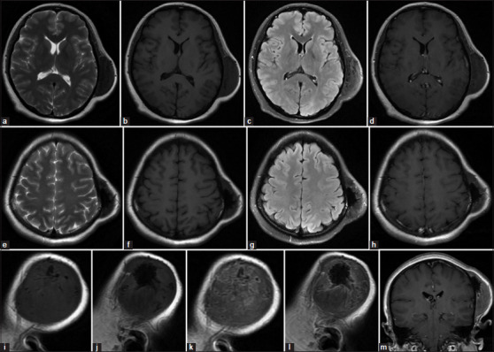
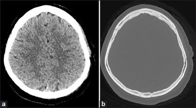
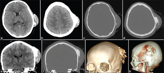
Desmoplastic fibroma (DF) is an uncommon benign bone tumor that typically affects the facial bones, with cerebral cranium involvement being extremely rare. We report a unique case of DF in the parietal bone of a 28-year-old woman, notable for its rapid growth during pregnancy-a phenomenon not previously documented. The imaging features of this case also differ from all but one previously reported case. The patient underwent surgical removal, and histopathology confirmed the diagnosis of DF (collagenous fibroma). After 17 months of follow-up, no local recurrence was observed. We also provide a comprehensive review of 32 cases involving DF of the cerebral cranium, analyzing clinical features, imaging findings, treatment methods, and recurrence patterns. This case highlights the importance of considering DF in the differential diagnosis of cranial lesions, particularly in pregnant patients with rapid tumor growth. Complete surgical resection with a wide margin remains the recommended treatment to minimize recurrence risk.
期刊介绍:
The Journal of Clinical Imaging Science (JCIS) is an open access peer-reviewed journal committed to publishing high-quality articles in the field of Imaging Science. The journal aims to present Imaging Science and relevant clinical information in an understandable and useful format. The journal is owned and published by the Scientific Scholar. Audience Our audience includes Radiologists, Researchers, Clinicians, medical professionals and students. Review process JCIS has a highly rigorous peer-review process that makes sure that manuscripts are scientifically accurate, relevant, novel and important. Authors disclose all conflicts, affiliations and financial associations such that the published content is not biased.

 求助内容:
求助内容: 应助结果提醒方式:


