{"title":"FAPI-04-PET/CT鉴别高级别胶质瘤复发及治疗后变化的初步评价","authors":"Indraja D Dev, Ameya D Puranik, Venkatesh Rangarajan, Sukriti Patra, Nilendu Purandare, Arpita Sahu, Amitkumar Choudhary, Kajari Bhattacharya, Tejpal Gupta, Abhishek Chatterjee, Archya Dasgupta, Aliasgar Moiyadi, Prakash Shetty, Vikas Singh, Epari Sridhar, Ayushi Sahay, Aekta Shah, Suchismita Ghosh, Sayak Choudhury, Sneha Shah, Archi Agrawal","doi":"10.37349/etat.2024.00276","DOIUrl":null,"url":null,"abstract":"<p><p>Fibroblast-activated protein (FAP) expression in glial cells is attributed to FAP-positive foci on tumor vessels and neoplastic cells. Preclinical and pilot studies have shown FAP expression in high-grade gliomas. We aimed at comparing PET imaging with FAP-inhibitor (FAPI-PET) with current standard, i.e., fluoro-ethyl tyrosine (FET) PET in post-treatment setting to differentiate recurrence and post-treatment changes. 6 patients with WHO Grade III and IV glioma who received standard treatment underwent Ga-68-FAPI-04 PET/CT (FAPI-PET/CT). Tracer uptake greater than background was considered positive. FET PET was performed and interpreted as per institutional standards, which formed the basis of treatment decision. There was concordance between FAPI expression and FET uptake in 5 patients suggestive of disease recurrence. There was no FAPI expression seen in 1 patient, in whom FET PET was suggestive of post-treatment changes. FAPI PET uptake correlated with amino acid expression to differentiate post treatment changes from recurrence in high-grade glial tumors; further validation with prospective study and histopathological confirmation is needed.</p>","PeriodicalId":73002,"journal":{"name":"Exploration of targeted anti-tumor therapy","volume":"5 6","pages":"1289-1296"},"PeriodicalIF":0.0000,"publicationDate":"2024-01-01","publicationTypes":"Journal Article","fieldsOfStudy":null,"isOpenAccess":false,"openAccessPdf":"https://www.ncbi.nlm.nih.gov/pmc/articles/PMC11700622/pdf/","citationCount":"0","resultStr":"{\"title\":\"Preliminary evaluation of FAPI-04-PET/CT for differentiating recurrence and post-treatment changes in high-grade gliomas.\",\"authors\":\"Indraja D Dev, Ameya D Puranik, Venkatesh Rangarajan, Sukriti Patra, Nilendu Purandare, Arpita Sahu, Amitkumar Choudhary, Kajari Bhattacharya, Tejpal Gupta, Abhishek Chatterjee, Archya Dasgupta, Aliasgar Moiyadi, Prakash Shetty, Vikas Singh, Epari Sridhar, Ayushi Sahay, Aekta Shah, Suchismita Ghosh, Sayak Choudhury, Sneha Shah, Archi Agrawal\",\"doi\":\"10.37349/etat.2024.00276\",\"DOIUrl\":null,\"url\":null,\"abstract\":\"<p><p>Fibroblast-activated protein (FAP) expression in glial cells is attributed to FAP-positive foci on tumor vessels and neoplastic cells. Preclinical and pilot studies have shown FAP expression in high-grade gliomas. We aimed at comparing PET imaging with FAP-inhibitor (FAPI-PET) with current standard, i.e., fluoro-ethyl tyrosine (FET) PET in post-treatment setting to differentiate recurrence and post-treatment changes. 6 patients with WHO Grade III and IV glioma who received standard treatment underwent Ga-68-FAPI-04 PET/CT (FAPI-PET/CT). Tracer uptake greater than background was considered positive. FET PET was performed and interpreted as per institutional standards, which formed the basis of treatment decision. There was concordance between FAPI expression and FET uptake in 5 patients suggestive of disease recurrence. There was no FAPI expression seen in 1 patient, in whom FET PET was suggestive of post-treatment changes. FAPI PET uptake correlated with amino acid expression to differentiate post treatment changes from recurrence in high-grade glial tumors; further validation with prospective study and histopathological confirmation is needed.</p>\",\"PeriodicalId\":73002,\"journal\":{\"name\":\"Exploration of targeted anti-tumor therapy\",\"volume\":\"5 6\",\"pages\":\"1289-1296\"},\"PeriodicalIF\":0.0000,\"publicationDate\":\"2024-01-01\",\"publicationTypes\":\"Journal Article\",\"fieldsOfStudy\":null,\"isOpenAccess\":false,\"openAccessPdf\":\"https://www.ncbi.nlm.nih.gov/pmc/articles/PMC11700622/pdf/\",\"citationCount\":\"0\",\"resultStr\":null,\"platform\":\"Semanticscholar\",\"paperid\":null,\"PeriodicalName\":\"Exploration of targeted anti-tumor therapy\",\"FirstCategoryId\":\"1085\",\"ListUrlMain\":\"https://doi.org/10.37349/etat.2024.00276\",\"RegionNum\":0,\"RegionCategory\":null,\"ArticlePicture\":[],\"TitleCN\":null,\"AbstractTextCN\":null,\"PMCID\":null,\"EPubDate\":\"2024/10/31 0:00:00\",\"PubModel\":\"Epub\",\"JCR\":\"Q3\",\"JCRName\":\"Medicine\",\"Score\":null,\"Total\":0}","platform":"Semanticscholar","paperid":null,"PeriodicalName":"Exploration of targeted anti-tumor therapy","FirstCategoryId":"1085","ListUrlMain":"https://doi.org/10.37349/etat.2024.00276","RegionNum":0,"RegionCategory":null,"ArticlePicture":[],"TitleCN":null,"AbstractTextCN":null,"PMCID":null,"EPubDate":"2024/10/31 0:00:00","PubModel":"Epub","JCR":"Q3","JCRName":"Medicine","Score":null,"Total":0}
Preliminary evaluation of FAPI-04-PET/CT for differentiating recurrence and post-treatment changes in high-grade gliomas.
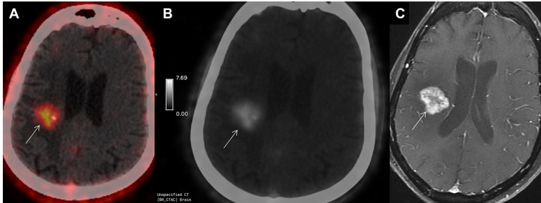
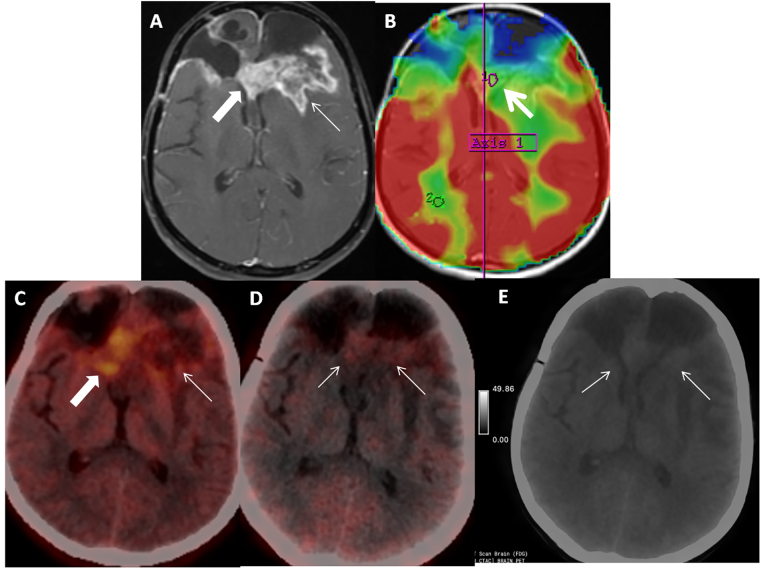
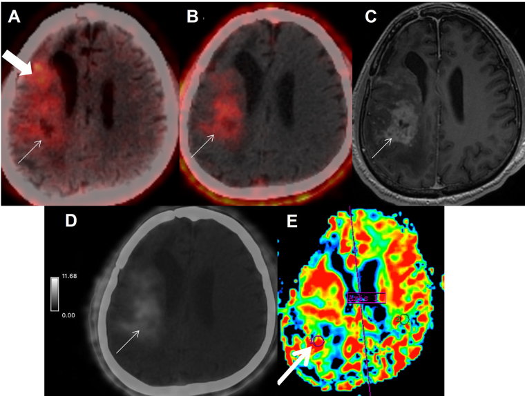
Fibroblast-activated protein (FAP) expression in glial cells is attributed to FAP-positive foci on tumor vessels and neoplastic cells. Preclinical and pilot studies have shown FAP expression in high-grade gliomas. We aimed at comparing PET imaging with FAP-inhibitor (FAPI-PET) with current standard, i.e., fluoro-ethyl tyrosine (FET) PET in post-treatment setting to differentiate recurrence and post-treatment changes. 6 patients with WHO Grade III and IV glioma who received standard treatment underwent Ga-68-FAPI-04 PET/CT (FAPI-PET/CT). Tracer uptake greater than background was considered positive. FET PET was performed and interpreted as per institutional standards, which formed the basis of treatment decision. There was concordance between FAPI expression and FET uptake in 5 patients suggestive of disease recurrence. There was no FAPI expression seen in 1 patient, in whom FET PET was suggestive of post-treatment changes. FAPI PET uptake correlated with amino acid expression to differentiate post treatment changes from recurrence in high-grade glial tumors; further validation with prospective study and histopathological confirmation is needed.

 求助内容:
求助内容: 应助结果提醒方式:


