David Shin , Ethan Vyhmeister , Daniel Im , Andrew Fay , Owen Faehner , Andrew Cabrera , Alexander Bouterse , Lauren Seo , Derran Bedward , Mei Carter , Davis Carter , Jacob Razzouk , Omar Ramos , Nathaniel Wycliffe , Wayne Cheng , Olumide Danisa
{"title":"腰椎间盘间隙高度与神经间孔尺寸和患者特征的关系:使用计算机断层扫描对L1-S1的形态计量学分析。","authors":"David Shin , Ethan Vyhmeister , Daniel Im , Andrew Fay , Owen Faehner , Andrew Cabrera , Alexander Bouterse , Lauren Seo , Derran Bedward , Mei Carter , Davis Carter , Jacob Razzouk , Omar Ramos , Nathaniel Wycliffe , Wayne Cheng , Olumide Danisa","doi":"10.1016/j.bas.2024.104162","DOIUrl":null,"url":null,"abstract":"<div><h3>Introduction</h3><div>The normative relationship between lumbar intervertebral disc space height (DSH) and neuroforaminal dimensions (NFD) has yet to be defined.</div></div><div><h3>Research question</h3><div>The purpose of this study was to investigate the relationship between lumbar DSH and NFD using computed tomography (CT), accounting for influences of patient demographic and anthropometric characteristics.</div></div><div><h3>Materials and methods</h3><div>We analyzed CT imaging of 350 female and 350 male patients. Anterior, middle, and posterior DSH were measured. NFD were defined as sagittal anterior-to-posterior (AP) width, axial AP width, foraminal height, and area. Statistical analyses were performed to assess associations among DSH, NFD, and patient height, weight, body mass index, sex, and ethnicity.</div></div><div><h3>Results</h3><div>Irrespective of disc level, mean anterior, middle, and posterior DSH were 7.98 mm (n = 3500), 8.16 mm (n = 3500), and 4.09 mm (n = 3500). DSH measurements demonstrated increasing, linear trends moving caudally from L1-L2 to L5-S1, while NFD demonstrated a unimodal distribution pattern with largest NFD at L3-L4 and smallest NFD at L1-2 and L5-S1. Male patients demonstrated larger DSH compared to female patients from L1-S1. Asian patients demonstrated taller DSH across all levels L1-S1.</div></div><div><h3>Discussion and conclusion</h3><div>This study describes 38,500 CT-based L1-S1 DSH and NFD in young patients without spinal pathology. DSH follows an increasing trend moving caudally from L1-S1, while NFD demonstrate a unimodal distribution clustered at L3-L4. NFD are not moderately or strongly associated with DSH. DSH is influenced by sex and ethnicity but is not moderately or strongly influenced by patient height, weight, and BMI.</div></div>","PeriodicalId":72443,"journal":{"name":"Brain & spine","volume":"5 ","pages":"Article 104162"},"PeriodicalIF":2.5000,"publicationDate":"2025-01-01","publicationTypes":"Journal Article","fieldsOfStudy":null,"isOpenAccess":false,"openAccessPdf":"https://www.ncbi.nlm.nih.gov/pmc/articles/PMC11700277/pdf/","citationCount":"0","resultStr":"{\"title\":\"Lumbar disc space height in relation to neural foraminal dimensions and patient characteristics: A morphometric analysis from L1-S1 using computed tomography\",\"authors\":\"David Shin , Ethan Vyhmeister , Daniel Im , Andrew Fay , Owen Faehner , Andrew Cabrera , Alexander Bouterse , Lauren Seo , Derran Bedward , Mei Carter , Davis Carter , Jacob Razzouk , Omar Ramos , Nathaniel Wycliffe , Wayne Cheng , Olumide Danisa\",\"doi\":\"10.1016/j.bas.2024.104162\",\"DOIUrl\":null,\"url\":null,\"abstract\":\"<div><h3>Introduction</h3><div>The normative relationship between lumbar intervertebral disc space height (DSH) and neuroforaminal dimensions (NFD) has yet to be defined.</div></div><div><h3>Research question</h3><div>The purpose of this study was to investigate the relationship between lumbar DSH and NFD using computed tomography (CT), accounting for influences of patient demographic and anthropometric characteristics.</div></div><div><h3>Materials and methods</h3><div>We analyzed CT imaging of 350 female and 350 male patients. Anterior, middle, and posterior DSH were measured. NFD were defined as sagittal anterior-to-posterior (AP) width, axial AP width, foraminal height, and area. Statistical analyses were performed to assess associations among DSH, NFD, and patient height, weight, body mass index, sex, and ethnicity.</div></div><div><h3>Results</h3><div>Irrespective of disc level, mean anterior, middle, and posterior DSH were 7.98 mm (n = 3500), 8.16 mm (n = 3500), and 4.09 mm (n = 3500). DSH measurements demonstrated increasing, linear trends moving caudally from L1-L2 to L5-S1, while NFD demonstrated a unimodal distribution pattern with largest NFD at L3-L4 and smallest NFD at L1-2 and L5-S1. Male patients demonstrated larger DSH compared to female patients from L1-S1. Asian patients demonstrated taller DSH across all levels L1-S1.</div></div><div><h3>Discussion and conclusion</h3><div>This study describes 38,500 CT-based L1-S1 DSH and NFD in young patients without spinal pathology. DSH follows an increasing trend moving caudally from L1-S1, while NFD demonstrate a unimodal distribution clustered at L3-L4. NFD are not moderately or strongly associated with DSH. DSH is influenced by sex and ethnicity but is not moderately or strongly influenced by patient height, weight, and BMI.</div></div>\",\"PeriodicalId\":72443,\"journal\":{\"name\":\"Brain & spine\",\"volume\":\"5 \",\"pages\":\"Article 104162\"},\"PeriodicalIF\":2.5000,\"publicationDate\":\"2025-01-01\",\"publicationTypes\":\"Journal Article\",\"fieldsOfStudy\":null,\"isOpenAccess\":false,\"openAccessPdf\":\"https://www.ncbi.nlm.nih.gov/pmc/articles/PMC11700277/pdf/\",\"citationCount\":\"0\",\"resultStr\":null,\"platform\":\"Semanticscholar\",\"paperid\":null,\"PeriodicalName\":\"Brain & spine\",\"FirstCategoryId\":\"1085\",\"ListUrlMain\":\"https://www.sciencedirect.com/science/article/pii/S2772529424014188\",\"RegionNum\":0,\"RegionCategory\":null,\"ArticlePicture\":[],\"TitleCN\":null,\"AbstractTextCN\":null,\"PMCID\":null,\"EPubDate\":\"\",\"PubModel\":\"\",\"JCR\":\"Q3\",\"JCRName\":\"CLINICAL NEUROLOGY\",\"Score\":null,\"Total\":0}","platform":"Semanticscholar","paperid":null,"PeriodicalName":"Brain & spine","FirstCategoryId":"1085","ListUrlMain":"https://www.sciencedirect.com/science/article/pii/S2772529424014188","RegionNum":0,"RegionCategory":null,"ArticlePicture":[],"TitleCN":null,"AbstractTextCN":null,"PMCID":null,"EPubDate":"","PubModel":"","JCR":"Q3","JCRName":"CLINICAL NEUROLOGY","Score":null,"Total":0}
引用次数: 0
摘要
导论:腰椎间盘间隙高度(DSH)与神经间孔尺寸(NFD)之间的规范关系尚未明确。研究问题:本研究的目的是利用计算机断层扫描(CT)研究腰椎DSH和NFD之间的关系,考虑患者人口统计学和人体测量学特征的影响。材料与方法:对350例女性和350例男性患者的CT影像进行分析。测量前、中、后DSH。NFD被定义为矢状面前后(AP)宽度、轴向AP宽度、椎间孔高度和面积。统计分析评估DSH、NFD与患者身高、体重、体重指数、性别和种族之间的关系。结果:无论椎间盘水平如何,平均前、中、后椎间盘突出度分别为7.98 mm (n = 3500)、8.16 mm (n = 3500)和4.09 mm (n = 3500)。DSH测量结果显示,从L1-L2到L5-S1呈线性增加趋势,而NFD呈单峰分布模式,最大NFD位于L3-L4,最小NFD位于L1-2和L5-S1。L1-S1期男性患者的DSH比女性患者大。亚洲患者在L1-S1所有水平均表现出较高的DSH。讨论与结论:本研究描述了38,500例无脊柱病理的年轻患者基于ct的L1-S1 DSH和NFD。DSH从L1-S1逐渐增加,而NFD在L3-L4呈单峰分布。NFD与DSH没有中度或强烈的相关性。DSH受性别和种族的影响,但不受患者身高、体重和BMI的中度或强烈影响。
Lumbar disc space height in relation to neural foraminal dimensions and patient characteristics: A morphometric analysis from L1-S1 using computed tomography
Introduction
The normative relationship between lumbar intervertebral disc space height (DSH) and neuroforaminal dimensions (NFD) has yet to be defined.
Research question
The purpose of this study was to investigate the relationship between lumbar DSH and NFD using computed tomography (CT), accounting for influences of patient demographic and anthropometric characteristics.
Materials and methods
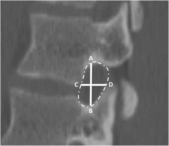
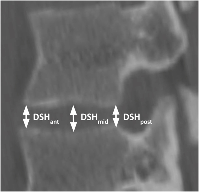
We analyzed CT imaging of 350 female and 350 male patients. Anterior, middle, and posterior DSH were measured. NFD were defined as sagittal anterior-to-posterior (AP) width, axial AP width, foraminal height, and area. Statistical analyses were performed to assess associations among DSH, NFD, and patient height, weight, body mass index, sex, and ethnicity.
Results
Irrespective of disc level, mean anterior, middle, and posterior DSH were 7.98 mm (n = 3500), 8.16 mm (n = 3500), and 4.09 mm (n = 3500). DSH measurements demonstrated increasing, linear trends moving caudally from L1-L2 to L5-S1, while NFD demonstrated a unimodal distribution pattern with largest NFD at L3-L4 and smallest NFD at L1-2 and L5-S1. Male patients demonstrated larger DSH compared to female patients from L1-S1. Asian patients demonstrated taller DSH across all levels L1-S1.
Discussion and conclusion
This study describes 38,500 CT-based L1-S1 DSH and NFD in young patients without spinal pathology. DSH follows an increasing trend moving caudally from L1-S1, while NFD demonstrate a unimodal distribution clustered at L3-L4. NFD are not moderately or strongly associated with DSH. DSH is influenced by sex and ethnicity but is not moderately or strongly influenced by patient height, weight, and BMI.

 求助内容:
求助内容: 应助结果提醒方式:


