Thao Thi Do, Lam Nguyen Le, Loc Truong Tan, Anh The Thien Dang, Duyen Ngoc Kim Huynh, My Hoan Truong, Luan Minh Nguyen
{"title":"锥形束CT对下颌椎管特征的探讨。","authors":"Thao Thi Do, Lam Nguyen Le, Loc Truong Tan, Anh The Thien Dang, Duyen Ngoc Kim Huynh, My Hoan Truong, Luan Minh Nguyen","doi":"10.4103/jos.jos_47_24","DOIUrl":null,"url":null,"abstract":"<p><strong>Context: </strong>The mandibular canal (MC) is an essential landmark that should be considered before any surgeries. Therefore, accurately assessing the location and characteristics of the MC in cone beam computed tomography (CBCT) imaging is very important.</p><p><strong>Aims: </strong>To determine the characteristics of the MC in relation to adjacent anatomical structures in CBCT projections.</p><p><strong>Settings and design: </strong>The convenience sampling method.</p><p><strong>Methods and material: </strong>This was a retrospective study of 112 CBCT images of Vietnamese patients aged 18 to 69 years, taken for clinical indications between 2018 and 2023. The evaluation was carried out by comparing and arranging the anatomical structures of different planes in three-dimensional space to assess and measure relevant dimensions.</p><p><strong>Statistical analysis used: </strong>Independent samples T-test.</p><p><strong>Results: </strong>The average diameter of the MC from the apex of the second premolar to the distal apex of the second molar, if there were no missing teeth in this segment, was 2.58 ± 0.52 mm (right) and 2.55 ± 0.54 mm (left). If there were a missing tooth in this segment, the measurements were 2.51 ± 0.79 mm (right) and 2.47 ± 0.45 mm (left). The difference between the two sides regarding the presence or absence of a missing tooth was not statistically significant.</p><p><strong>Conclusions: </strong>The precise localization of the MC related to the tooth apex and the diameter of the MC can vary in each person. CBCT indications should be considered when establishing treatment planning to avoid damaging the inferior alveolar nerve in the MC.</p>","PeriodicalId":16604,"journal":{"name":"Journal of Orthodontic Science","volume":"13 ","pages":"45"},"PeriodicalIF":0.0000,"publicationDate":"2024-11-25","publicationTypes":"Journal Article","fieldsOfStudy":null,"isOpenAccess":false,"openAccessPdf":"https://www.ncbi.nlm.nih.gov/pmc/articles/PMC11698245/pdf/","citationCount":"0","resultStr":"{\"title\":\"Investigating the characteristics of the mandibular canal in cone beam CT.\",\"authors\":\"Thao Thi Do, Lam Nguyen Le, Loc Truong Tan, Anh The Thien Dang, Duyen Ngoc Kim Huynh, My Hoan Truong, Luan Minh Nguyen\",\"doi\":\"10.4103/jos.jos_47_24\",\"DOIUrl\":null,\"url\":null,\"abstract\":\"<p><strong>Context: </strong>The mandibular canal (MC) is an essential landmark that should be considered before any surgeries. Therefore, accurately assessing the location and characteristics of the MC in cone beam computed tomography (CBCT) imaging is very important.</p><p><strong>Aims: </strong>To determine the characteristics of the MC in relation to adjacent anatomical structures in CBCT projections.</p><p><strong>Settings and design: </strong>The convenience sampling method.</p><p><strong>Methods and material: </strong>This was a retrospective study of 112 CBCT images of Vietnamese patients aged 18 to 69 years, taken for clinical indications between 2018 and 2023. The evaluation was carried out by comparing and arranging the anatomical structures of different planes in three-dimensional space to assess and measure relevant dimensions.</p><p><strong>Statistical analysis used: </strong>Independent samples T-test.</p><p><strong>Results: </strong>The average diameter of the MC from the apex of the second premolar to the distal apex of the second molar, if there were no missing teeth in this segment, was 2.58 ± 0.52 mm (right) and 2.55 ± 0.54 mm (left). If there were a missing tooth in this segment, the measurements were 2.51 ± 0.79 mm (right) and 2.47 ± 0.45 mm (left). The difference between the two sides regarding the presence or absence of a missing tooth was not statistically significant.</p><p><strong>Conclusions: </strong>The precise localization of the MC related to the tooth apex and the diameter of the MC can vary in each person. CBCT indications should be considered when establishing treatment planning to avoid damaging the inferior alveolar nerve in the MC.</p>\",\"PeriodicalId\":16604,\"journal\":{\"name\":\"Journal of Orthodontic Science\",\"volume\":\"13 \",\"pages\":\"45\"},\"PeriodicalIF\":0.0000,\"publicationDate\":\"2024-11-25\",\"publicationTypes\":\"Journal Article\",\"fieldsOfStudy\":null,\"isOpenAccess\":false,\"openAccessPdf\":\"https://www.ncbi.nlm.nih.gov/pmc/articles/PMC11698245/pdf/\",\"citationCount\":\"0\",\"resultStr\":null,\"platform\":\"Semanticscholar\",\"paperid\":null,\"PeriodicalName\":\"Journal of Orthodontic Science\",\"FirstCategoryId\":\"1085\",\"ListUrlMain\":\"https://doi.org/10.4103/jos.jos_47_24\",\"RegionNum\":0,\"RegionCategory\":null,\"ArticlePicture\":[],\"TitleCN\":null,\"AbstractTextCN\":null,\"PMCID\":null,\"EPubDate\":\"2024/1/1 0:00:00\",\"PubModel\":\"eCollection\",\"JCR\":\"Q2\",\"JCRName\":\"Dentistry\",\"Score\":null,\"Total\":0}","platform":"Semanticscholar","paperid":null,"PeriodicalName":"Journal of Orthodontic Science","FirstCategoryId":"1085","ListUrlMain":"https://doi.org/10.4103/jos.jos_47_24","RegionNum":0,"RegionCategory":null,"ArticlePicture":[],"TitleCN":null,"AbstractTextCN":null,"PMCID":null,"EPubDate":"2024/1/1 0:00:00","PubModel":"eCollection","JCR":"Q2","JCRName":"Dentistry","Score":null,"Total":0}
Investigating the characteristics of the mandibular canal in cone beam CT.
Context: The mandibular canal (MC) is an essential landmark that should be considered before any surgeries. Therefore, accurately assessing the location and characteristics of the MC in cone beam computed tomography (CBCT) imaging is very important.
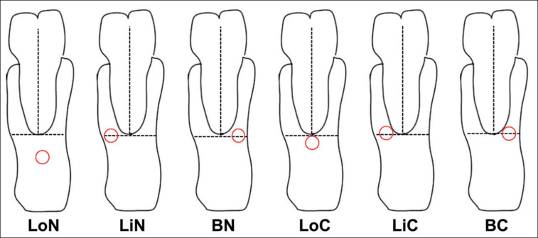
Aims: To determine the characteristics of the MC in relation to adjacent anatomical structures in CBCT projections.
Settings and design: The convenience sampling method.
Methods and material: This was a retrospective study of 112 CBCT images of Vietnamese patients aged 18 to 69 years, taken for clinical indications between 2018 and 2023. The evaluation was carried out by comparing and arranging the anatomical structures of different planes in three-dimensional space to assess and measure relevant dimensions.
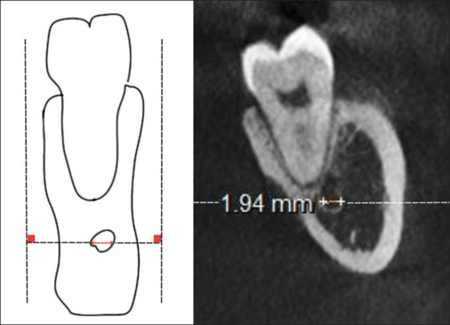
Results: The average diameter of the MC from the apex of the second premolar to the distal apex of the second molar, if there were no missing teeth in this segment, was 2.58 ± 0.52 mm (right) and 2.55 ± 0.54 mm (left). If there were a missing tooth in this segment, the measurements were 2.51 ± 0.79 mm (right) and 2.47 ± 0.45 mm (left). The difference between the two sides regarding the presence or absence of a missing tooth was not statistically significant.
Conclusions: The precise localization of the MC related to the tooth apex and the diameter of the MC can vary in each person. CBCT indications should be considered when establishing treatment planning to avoid damaging the inferior alveolar nerve in the MC.


 求助内容:
求助内容: 应助结果提醒方式:


