Amgad Droby, Avital Yoffe-Vasiliev, Daniel Atias, Kyle B. Fraser, Omar S. Mabrouk, Nurit Omer, Anat Bar-Shira, Mali Gana-Weisz, Orly Goldstein, Moran Artzi, Dafna Ben Bashat, Roy N. Alcalay, Avi Orr-Urtreger, Julia C. Shirvan, Jesse M. Cedarbaum, Nir Giladi, Anat Mirelman, Avner Thaler
{"title":"帕金森病患者脑脊液α-突触核蛋白聚集的影像学标志物","authors":"Amgad Droby, Avital Yoffe-Vasiliev, Daniel Atias, Kyle B. Fraser, Omar S. Mabrouk, Nurit Omer, Anat Bar-Shira, Mali Gana-Weisz, Orly Goldstein, Moran Artzi, Dafna Ben Bashat, Roy N. Alcalay, Avi Orr-Urtreger, Julia C. Shirvan, Jesse M. Cedarbaum, Nir Giladi, Anat Mirelman, Avner Thaler","doi":"10.1038/s41531-024-00854-4","DOIUrl":null,"url":null,"abstract":"<p>Alpha-synuclein (αS) aggregation is a widely regarded hallmark of Parkinson’s disease (PD) and can be detected through synuclein amplification assays (SAA). This study investigated the association between cerebrospinal fluid (CSF) radiological measures in 41 PD patients (14 iPD, 14 <i>GBA1</i>-PD, 13 <i>LRRK2</i>-PD) and 14 age-and-sex-matched healthy controls. Quantitative measures including striatal binding ratios (SBR), whole-brain and deep gray matter volumes, neuromelanin-MRI (NM-MRI), functional connectivity (FC), and white matter (WM) diffusion-tensor imaging (DTI) were calculated. Nine <i>LRRK2</i>-PD patients were SAA-negative (PD-SAA−). PD-SAA+ patients showed lower whole-brain gray matter, putamenal, brainstem, and substantia nigra volumes, reduced FC in the left caudate, and lower fractional anisotropy in the left fronto-occipital fasciculus compared to PD-SAA−. Taken together, αS aggregation was observed in iPD, <i>GBA1</i>-PD, and 38% of <i>LRRK2</i>-PD patients, and this was associated with reduced regional brain volumes, altered caudal FC, and SBRs. These changes were less pronounced in PD-SAA−, possibly suggesting a milder neurodegenerative process.</p>","PeriodicalId":19706,"journal":{"name":"NPJ Parkinson's Disease","volume":"19 1","pages":""},"PeriodicalIF":6.7000,"publicationDate":"2025-01-03","publicationTypes":"Journal Article","fieldsOfStudy":null,"isOpenAccess":false,"openAccessPdf":"","citationCount":"0","resultStr":"{\"title\":\"Radiological markers of CSF α-synuclein aggregation in Parkinson’s disease patients\",\"authors\":\"Amgad Droby, Avital Yoffe-Vasiliev, Daniel Atias, Kyle B. Fraser, Omar S. Mabrouk, Nurit Omer, Anat Bar-Shira, Mali Gana-Weisz, Orly Goldstein, Moran Artzi, Dafna Ben Bashat, Roy N. Alcalay, Avi Orr-Urtreger, Julia C. Shirvan, Jesse M. Cedarbaum, Nir Giladi, Anat Mirelman, Avner Thaler\",\"doi\":\"10.1038/s41531-024-00854-4\",\"DOIUrl\":null,\"url\":null,\"abstract\":\"<p>Alpha-synuclein (αS) aggregation is a widely regarded hallmark of Parkinson’s disease (PD) and can be detected through synuclein amplification assays (SAA). This study investigated the association between cerebrospinal fluid (CSF) radiological measures in 41 PD patients (14 iPD, 14 <i>GBA1</i>-PD, 13 <i>LRRK2</i>-PD) and 14 age-and-sex-matched healthy controls. Quantitative measures including striatal binding ratios (SBR), whole-brain and deep gray matter volumes, neuromelanin-MRI (NM-MRI), functional connectivity (FC), and white matter (WM) diffusion-tensor imaging (DTI) were calculated. Nine <i>LRRK2</i>-PD patients were SAA-negative (PD-SAA−). PD-SAA+ patients showed lower whole-brain gray matter, putamenal, brainstem, and substantia nigra volumes, reduced FC in the left caudate, and lower fractional anisotropy in the left fronto-occipital fasciculus compared to PD-SAA−. Taken together, αS aggregation was observed in iPD, <i>GBA1</i>-PD, and 38% of <i>LRRK2</i>-PD patients, and this was associated with reduced regional brain volumes, altered caudal FC, and SBRs. These changes were less pronounced in PD-SAA−, possibly suggesting a milder neurodegenerative process.</p>\",\"PeriodicalId\":19706,\"journal\":{\"name\":\"NPJ Parkinson's Disease\",\"volume\":\"19 1\",\"pages\":\"\"},\"PeriodicalIF\":6.7000,\"publicationDate\":\"2025-01-03\",\"publicationTypes\":\"Journal Article\",\"fieldsOfStudy\":null,\"isOpenAccess\":false,\"openAccessPdf\":\"\",\"citationCount\":\"0\",\"resultStr\":null,\"platform\":\"Semanticscholar\",\"paperid\":null,\"PeriodicalName\":\"NPJ Parkinson's Disease\",\"FirstCategoryId\":\"3\",\"ListUrlMain\":\"https://doi.org/10.1038/s41531-024-00854-4\",\"RegionNum\":1,\"RegionCategory\":\"医学\",\"ArticlePicture\":[],\"TitleCN\":null,\"AbstractTextCN\":null,\"PMCID\":null,\"EPubDate\":\"\",\"PubModel\":\"\",\"JCR\":\"Q1\",\"JCRName\":\"NEUROSCIENCES\",\"Score\":null,\"Total\":0}","platform":"Semanticscholar","paperid":null,"PeriodicalName":"NPJ Parkinson's Disease","FirstCategoryId":"3","ListUrlMain":"https://doi.org/10.1038/s41531-024-00854-4","RegionNum":1,"RegionCategory":"医学","ArticlePicture":[],"TitleCN":null,"AbstractTextCN":null,"PMCID":null,"EPubDate":"","PubModel":"","JCR":"Q1","JCRName":"NEUROSCIENCES","Score":null,"Total":0}
Radiological markers of CSF α-synuclein aggregation in Parkinson’s disease patients
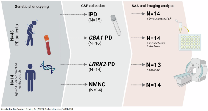
Alpha-synuclein (αS) aggregation is a widely regarded hallmark of Parkinson’s disease (PD) and can be detected through synuclein amplification assays (SAA). This study investigated the association between cerebrospinal fluid (CSF) radiological measures in 41 PD patients (14 iPD, 14 GBA1-PD, 13 LRRK2-PD) and 14 age-and-sex-matched healthy controls. Quantitative measures including striatal binding ratios (SBR), whole-brain and deep gray matter volumes, neuromelanin-MRI (NM-MRI), functional connectivity (FC), and white matter (WM) diffusion-tensor imaging (DTI) were calculated. Nine LRRK2-PD patients were SAA-negative (PD-SAA−). PD-SAA+ patients showed lower whole-brain gray matter, putamenal, brainstem, and substantia nigra volumes, reduced FC in the left caudate, and lower fractional anisotropy in the left fronto-occipital fasciculus compared to PD-SAA−. Taken together, αS aggregation was observed in iPD, GBA1-PD, and 38% of LRRK2-PD patients, and this was associated with reduced regional brain volumes, altered caudal FC, and SBRs. These changes were less pronounced in PD-SAA−, possibly suggesting a milder neurodegenerative process.
期刊介绍:
npj Parkinson's Disease is a comprehensive open access journal that covers a wide range of research areas related to Parkinson's disease. It publishes original studies in basic science, translational research, and clinical investigations. The journal is dedicated to advancing our understanding of Parkinson's disease by exploring various aspects such as anatomy, etiology, genetics, cellular and molecular physiology, neurophysiology, epidemiology, and therapeutic development. By providing free and immediate access to the scientific and Parkinson's disease community, npj Parkinson's Disease promotes collaboration and knowledge sharing among researchers and healthcare professionals.
 求助内容:
求助内容: 应助结果提醒方式:


