{"title":"房颤消融继发肺静脉狭窄的通气/灌注失配。","authors":"Leah Anne Christine L Bollos, Ryosuke Kasai, Hideki Otsuka, Yoichi Otomi, Tomomi Matsuura, Tamaki Otani, Koji Yamaguchi, Takanori Bando, Yuya Ueki, Noritake Matsuda, Satoru Takashi, Shota Azane, Yamato Kunikane, Shoichiro Takao, Shusuke Yagi, Masataka Sata, Hitoshi Ikushima, Masafumi Harada","doi":"10.22038/aojnmb.2024.79650.1561","DOIUrl":null,"url":null,"abstract":"<p><p>We present two patients with a history of paroxysmal atrial fibrillation who developed pulmonary vein stenosis (PVS) following atrial fibrillation (AF) ablation. Case 1 involved a female patient in her 50s who was asymptomatic for pulmonary symptoms but was found to have a high degree of left superior PVS 15 months after AF ablation. This was demonstrated using contrast-enhanced computed tomography (CE-CT) and supported by findings of perfusion defects on ventilation-perfusion (V/Q) scan. Case 2 was a male patient in his 60s who developed progressive left superior PVS nine months after AF ablation, evidenced by serial CE-CT and V/Q scans. PVS is a rare but well-known complication of pulmonary vein ablation for the treatment of AF that can lead to severe complications if left untreated. V/Q scans effectively assess the functional significance of PVS by detecting abnormal blood flow segments. Although a V/Q mismatch characterized by reduced perfusion defects is more commonly used in evaluating pulmonary embolism, PVS should not be disregarded as a differential diagnosis. Few studies emphasize the utility of V/Q scans in managing PVS and highlight V/Q mismatch as a notable finding. This case report aimed to highlight their significance.</p>","PeriodicalId":8503,"journal":{"name":"Asia Oceania Journal of Nuclear Medicine and Biology","volume":"13 1","pages":"62-69"},"PeriodicalIF":0.0000,"publicationDate":"2025-01-01","publicationTypes":"Journal Article","fieldsOfStudy":null,"isOpenAccess":false,"openAccessPdf":"https://www.ncbi.nlm.nih.gov/pmc/articles/PMC11682475/pdf/","citationCount":"0","resultStr":"{\"title\":\"Ventilation/Perfusion Mismatch in Pulmonary Vein Stenosis Secondary to Atrial Fibrillation Ablation.\",\"authors\":\"Leah Anne Christine L Bollos, Ryosuke Kasai, Hideki Otsuka, Yoichi Otomi, Tomomi Matsuura, Tamaki Otani, Koji Yamaguchi, Takanori Bando, Yuya Ueki, Noritake Matsuda, Satoru Takashi, Shota Azane, Yamato Kunikane, Shoichiro Takao, Shusuke Yagi, Masataka Sata, Hitoshi Ikushima, Masafumi Harada\",\"doi\":\"10.22038/aojnmb.2024.79650.1561\",\"DOIUrl\":null,\"url\":null,\"abstract\":\"<p><p>We present two patients with a history of paroxysmal atrial fibrillation who developed pulmonary vein stenosis (PVS) following atrial fibrillation (AF) ablation. Case 1 involved a female patient in her 50s who was asymptomatic for pulmonary symptoms but was found to have a high degree of left superior PVS 15 months after AF ablation. This was demonstrated using contrast-enhanced computed tomography (CE-CT) and supported by findings of perfusion defects on ventilation-perfusion (V/Q) scan. Case 2 was a male patient in his 60s who developed progressive left superior PVS nine months after AF ablation, evidenced by serial CE-CT and V/Q scans. PVS is a rare but well-known complication of pulmonary vein ablation for the treatment of AF that can lead to severe complications if left untreated. V/Q scans effectively assess the functional significance of PVS by detecting abnormal blood flow segments. Although a V/Q mismatch characterized by reduced perfusion defects is more commonly used in evaluating pulmonary embolism, PVS should not be disregarded as a differential diagnosis. Few studies emphasize the utility of V/Q scans in managing PVS and highlight V/Q mismatch as a notable finding. This case report aimed to highlight their significance.</p>\",\"PeriodicalId\":8503,\"journal\":{\"name\":\"Asia Oceania Journal of Nuclear Medicine and Biology\",\"volume\":\"13 1\",\"pages\":\"62-69\"},\"PeriodicalIF\":0.0000,\"publicationDate\":\"2025-01-01\",\"publicationTypes\":\"Journal Article\",\"fieldsOfStudy\":null,\"isOpenAccess\":false,\"openAccessPdf\":\"https://www.ncbi.nlm.nih.gov/pmc/articles/PMC11682475/pdf/\",\"citationCount\":\"0\",\"resultStr\":null,\"platform\":\"Semanticscholar\",\"paperid\":null,\"PeriodicalName\":\"Asia Oceania Journal of Nuclear Medicine and Biology\",\"FirstCategoryId\":\"1085\",\"ListUrlMain\":\"https://doi.org/10.22038/aojnmb.2024.79650.1561\",\"RegionNum\":0,\"RegionCategory\":null,\"ArticlePicture\":[],\"TitleCN\":null,\"AbstractTextCN\":null,\"PMCID\":null,\"EPubDate\":\"\",\"PubModel\":\"\",\"JCR\":\"Q3\",\"JCRName\":\"Medicine\",\"Score\":null,\"Total\":0}","platform":"Semanticscholar","paperid":null,"PeriodicalName":"Asia Oceania Journal of Nuclear Medicine and Biology","FirstCategoryId":"1085","ListUrlMain":"https://doi.org/10.22038/aojnmb.2024.79650.1561","RegionNum":0,"RegionCategory":null,"ArticlePicture":[],"TitleCN":null,"AbstractTextCN":null,"PMCID":null,"EPubDate":"","PubModel":"","JCR":"Q3","JCRName":"Medicine","Score":null,"Total":0}
Ventilation/Perfusion Mismatch in Pulmonary Vein Stenosis Secondary to Atrial Fibrillation Ablation.
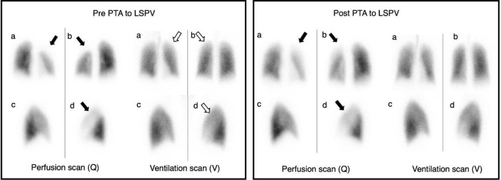
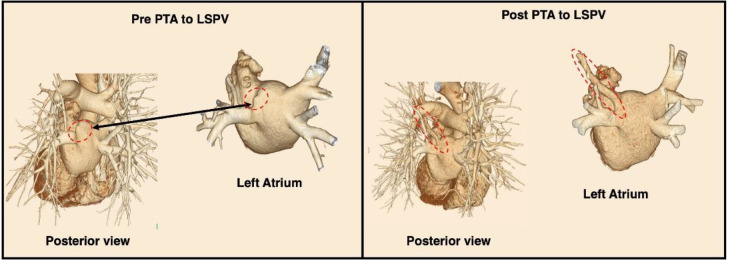
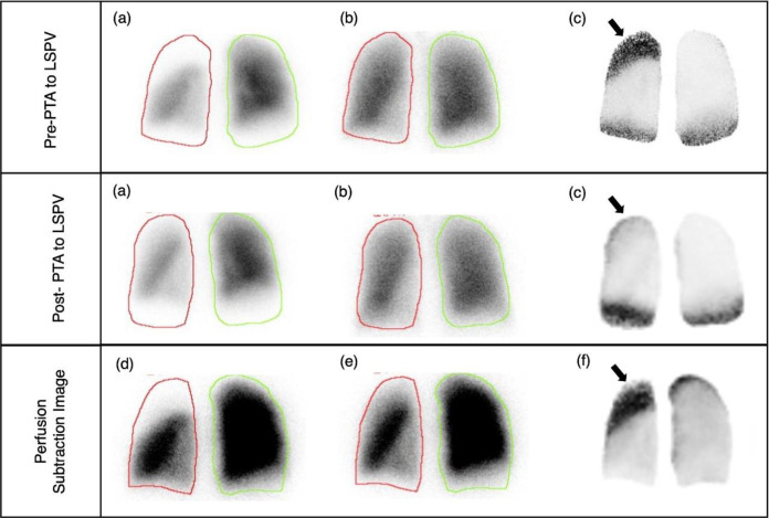
We present two patients with a history of paroxysmal atrial fibrillation who developed pulmonary vein stenosis (PVS) following atrial fibrillation (AF) ablation. Case 1 involved a female patient in her 50s who was asymptomatic for pulmonary symptoms but was found to have a high degree of left superior PVS 15 months after AF ablation. This was demonstrated using contrast-enhanced computed tomography (CE-CT) and supported by findings of perfusion defects on ventilation-perfusion (V/Q) scan. Case 2 was a male patient in his 60s who developed progressive left superior PVS nine months after AF ablation, evidenced by serial CE-CT and V/Q scans. PVS is a rare but well-known complication of pulmonary vein ablation for the treatment of AF that can lead to severe complications if left untreated. V/Q scans effectively assess the functional significance of PVS by detecting abnormal blood flow segments. Although a V/Q mismatch characterized by reduced perfusion defects is more commonly used in evaluating pulmonary embolism, PVS should not be disregarded as a differential diagnosis. Few studies emphasize the utility of V/Q scans in managing PVS and highlight V/Q mismatch as a notable finding. This case report aimed to highlight their significance.

 求助内容:
求助内容: 应助结果提醒方式:


