{"title":"18F-FDG俯伏乳腺硅光电倍增管PET/CT成像小病灶图像质量的改善","authors":"Nobuhiro Yada, Hiroyuki Kuroda, Toshihiko Kawamura, Mizuki Fukuda, Yoshinori Miyahara, Takeshi Yoshizako, Yasushi Kaji","doi":"10.22038/aojnmb.2024.78080.1553","DOIUrl":null,"url":null,"abstract":"<p><strong>Objectives: </strong>We investigated image quality and standardized uptake values (SUVs) for different lesion sizes using clinical data generated by <sup>18</sup>F-FDG-prone breast silicon photomultiplier (SiPM)-based positron emission tomography/computed tomography (PET/CT).</p><p><strong>Methods: </strong>We evaluated the effect of point-spread function (PSF) modeling and Gaussian filtering (Gau) and determined the optimal reconstruction conditions. We compared the signal-to-noise ratio (SNR), contrast, %coefficient of variation (%CV), SUV, and Likert scale score between ordered-subset expectation maximization (OSEM) time-of-flight (TOF) and OSEM+TOF+PSF in phantom and clinical studies. The conventional image was generated with OSEM+TOF_Gau 6 mm. The National Electrical Manufacturers Association body phantom with 10-mm hot sphere data was acquired for 5 min. Twenty-six patients (40 lesions, ranging from 3.7 to 63.0 mm) were examined using prone breast PET/CT with a breast positioner for breast cancer staging. PET data were acquired 125±9.7 min after intravenous injection of 220±16.1 MBq at 5 min/bed.</p><p><strong>Results: </strong>In the phantom study, a high SNR was obtained from a 3- to 5-mm Gaussian filter for OSEM+TOF+PSF. The contrast obtained with OSEM+TOF without Gaussian filtering was superior to that obtained with OSEM+TOF+PSF_Gau 4 mm. In the clinical study, the image quality depended on lesion size. The average SNR was significantly higher at 40.8% for lesions >20 mm with OSEM+TOF_Gau 6 mm than with OSEM+TOF without Gaussian filtering. The average contrast for lesions ≤10 mm was significantly higher by 42.0% with OSEM+TOF without Gaussian filtering than with OSEM+TOF_Gau 6 mm. The average SUV<sub>max</sub> of OSEM+TOF without Gaussian filtering significantly increased by 53.3% for lesions ≤10 mm.</p><p><strong>Conclusion: </strong>OSEM+TOF without Gaussian filtering provided good contrast and quantitative value for small lesions.</p>","PeriodicalId":8503,"journal":{"name":"Asia Oceania Journal of Nuclear Medicine and Biology","volume":"13 1","pages":"77-86"},"PeriodicalIF":0.0000,"publicationDate":"2025-01-01","publicationTypes":"Journal Article","fieldsOfStudy":null,"isOpenAccess":false,"openAccessPdf":"https://www.ncbi.nlm.nih.gov/pmc/articles/PMC11682473/pdf/","citationCount":"0","resultStr":"{\"title\":\"Improvement of image quality for small lesion sizes in <sup>18</sup>F-FDG prone breast silicon photomultiplier-based PET/CT imaging.\",\"authors\":\"Nobuhiro Yada, Hiroyuki Kuroda, Toshihiko Kawamura, Mizuki Fukuda, Yoshinori Miyahara, Takeshi Yoshizako, Yasushi Kaji\",\"doi\":\"10.22038/aojnmb.2024.78080.1553\",\"DOIUrl\":null,\"url\":null,\"abstract\":\"<p><strong>Objectives: </strong>We investigated image quality and standardized uptake values (SUVs) for different lesion sizes using clinical data generated by <sup>18</sup>F-FDG-prone breast silicon photomultiplier (SiPM)-based positron emission tomography/computed tomography (PET/CT).</p><p><strong>Methods: </strong>We evaluated the effect of point-spread function (PSF) modeling and Gaussian filtering (Gau) and determined the optimal reconstruction conditions. We compared the signal-to-noise ratio (SNR), contrast, %coefficient of variation (%CV), SUV, and Likert scale score between ordered-subset expectation maximization (OSEM) time-of-flight (TOF) and OSEM+TOF+PSF in phantom and clinical studies. The conventional image was generated with OSEM+TOF_Gau 6 mm. The National Electrical Manufacturers Association body phantom with 10-mm hot sphere data was acquired for 5 min. Twenty-six patients (40 lesions, ranging from 3.7 to 63.0 mm) were examined using prone breast PET/CT with a breast positioner for breast cancer staging. PET data were acquired 125±9.7 min after intravenous injection of 220±16.1 MBq at 5 min/bed.</p><p><strong>Results: </strong>In the phantom study, a high SNR was obtained from a 3- to 5-mm Gaussian filter for OSEM+TOF+PSF. The contrast obtained with OSEM+TOF without Gaussian filtering was superior to that obtained with OSEM+TOF+PSF_Gau 4 mm. In the clinical study, the image quality depended on lesion size. The average SNR was significantly higher at 40.8% for lesions >20 mm with OSEM+TOF_Gau 6 mm than with OSEM+TOF without Gaussian filtering. The average contrast for lesions ≤10 mm was significantly higher by 42.0% with OSEM+TOF without Gaussian filtering than with OSEM+TOF_Gau 6 mm. The average SUV<sub>max</sub> of OSEM+TOF without Gaussian filtering significantly increased by 53.3% for lesions ≤10 mm.</p><p><strong>Conclusion: </strong>OSEM+TOF without Gaussian filtering provided good contrast and quantitative value for small lesions.</p>\",\"PeriodicalId\":8503,\"journal\":{\"name\":\"Asia Oceania Journal of Nuclear Medicine and Biology\",\"volume\":\"13 1\",\"pages\":\"77-86\"},\"PeriodicalIF\":0.0000,\"publicationDate\":\"2025-01-01\",\"publicationTypes\":\"Journal Article\",\"fieldsOfStudy\":null,\"isOpenAccess\":false,\"openAccessPdf\":\"https://www.ncbi.nlm.nih.gov/pmc/articles/PMC11682473/pdf/\",\"citationCount\":\"0\",\"resultStr\":null,\"platform\":\"Semanticscholar\",\"paperid\":null,\"PeriodicalName\":\"Asia Oceania Journal of Nuclear Medicine and Biology\",\"FirstCategoryId\":\"1085\",\"ListUrlMain\":\"https://doi.org/10.22038/aojnmb.2024.78080.1553\",\"RegionNum\":0,\"RegionCategory\":null,\"ArticlePicture\":[],\"TitleCN\":null,\"AbstractTextCN\":null,\"PMCID\":null,\"EPubDate\":\"\",\"PubModel\":\"\",\"JCR\":\"Q3\",\"JCRName\":\"Medicine\",\"Score\":null,\"Total\":0}","platform":"Semanticscholar","paperid":null,"PeriodicalName":"Asia Oceania Journal of Nuclear Medicine and Biology","FirstCategoryId":"1085","ListUrlMain":"https://doi.org/10.22038/aojnmb.2024.78080.1553","RegionNum":0,"RegionCategory":null,"ArticlePicture":[],"TitleCN":null,"AbstractTextCN":null,"PMCID":null,"EPubDate":"","PubModel":"","JCR":"Q3","JCRName":"Medicine","Score":null,"Total":0}
Improvement of image quality for small lesion sizes in 18F-FDG prone breast silicon photomultiplier-based PET/CT imaging.
Objectives: We investigated image quality and standardized uptake values (SUVs) for different lesion sizes using clinical data generated by 18F-FDG-prone breast silicon photomultiplier (SiPM)-based positron emission tomography/computed tomography (PET/CT).
Methods: We evaluated the effect of point-spread function (PSF) modeling and Gaussian filtering (Gau) and determined the optimal reconstruction conditions. We compared the signal-to-noise ratio (SNR), contrast, %coefficient of variation (%CV), SUV, and Likert scale score between ordered-subset expectation maximization (OSEM) time-of-flight (TOF) and OSEM+TOF+PSF in phantom and clinical studies. The conventional image was generated with OSEM+TOF_Gau 6 mm. The National Electrical Manufacturers Association body phantom with 10-mm hot sphere data was acquired for 5 min. Twenty-six patients (40 lesions, ranging from 3.7 to 63.0 mm) were examined using prone breast PET/CT with a breast positioner for breast cancer staging. PET data were acquired 125±9.7 min after intravenous injection of 220±16.1 MBq at 5 min/bed.
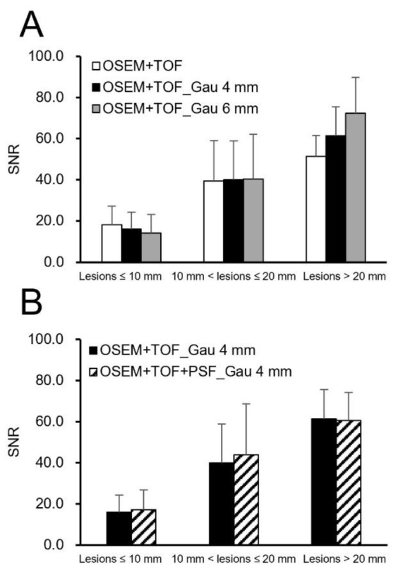
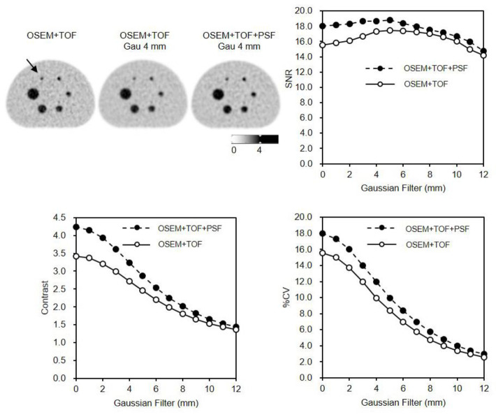
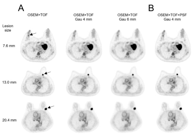
Results: In the phantom study, a high SNR was obtained from a 3- to 5-mm Gaussian filter for OSEM+TOF+PSF. The contrast obtained with OSEM+TOF without Gaussian filtering was superior to that obtained with OSEM+TOF+PSF_Gau 4 mm. In the clinical study, the image quality depended on lesion size. The average SNR was significantly higher at 40.8% for lesions >20 mm with OSEM+TOF_Gau 6 mm than with OSEM+TOF without Gaussian filtering. The average contrast for lesions ≤10 mm was significantly higher by 42.0% with OSEM+TOF without Gaussian filtering than with OSEM+TOF_Gau 6 mm. The average SUVmax of OSEM+TOF without Gaussian filtering significantly increased by 53.3% for lesions ≤10 mm.
Conclusion: OSEM+TOF without Gaussian filtering provided good contrast and quantitative value for small lesions.

 求助内容:
求助内容: 应助结果提醒方式:


