{"title":"18F-FDG PET/CT在淋巴管肉瘤中的表现:1例报告及文献复习。","authors":"Nitin Gupta","doi":"10.22038/aojnmb.2024.77689.1548","DOIUrl":null,"url":null,"abstract":"<p><strong>Objectives: </strong>Lymphangiosarcoma is a rare tumor that affects the upper limbs of patients who have undergone breast cancer surgery, including axillary dissection, followed by radiation therapy (RT) to the axilla and has a poor prognosis. <sup>18</sup>F-FDG PET/CT may enable the earlier detection of malignant transformation in a setting of chronic lymphedema and help evaluate the extent and staging of the tumor, allowing earlier initiation of treatment options.</p><p><strong>Case presentation: </strong>We herein report a case of cutaneous lymphangiosarcoma in a 47-year-old breast carcinoma patient, which occurred 9 years after initial surgery and radiation therapy. Distant metastases were detected on <sup>18</sup>F-FDG PET/CT. The patient underwent fore-quarter amputation of the upper limb and concurrent chemo-radiation therapy. However, she succumbed to her disease after 3 cycles of chemotherapy.</p><p><strong>Conclusions: </strong><sup>18</sup>F-FDG PET/CT scan helps in the early detection of malignant transformation and lymphangiosarcoma in a setting of chronic lymphedema in breast carcinoma patients following radiation therapy to the axilla. Furthermore, it helps determine the extent of regional spread and detect metastatic involvement, thus enabling better clinical management of these patients.</p>","PeriodicalId":8503,"journal":{"name":"Asia Oceania Journal of Nuclear Medicine and Biology","volume":"13 1","pages":"107-113"},"PeriodicalIF":0.0000,"publicationDate":"2025-01-01","publicationTypes":"Journal Article","fieldsOfStudy":null,"isOpenAccess":false,"openAccessPdf":"https://www.ncbi.nlm.nih.gov/pmc/articles/PMC11682481/pdf/","citationCount":"0","resultStr":"{\"title\":\"<sup>18</sup>F-FDG PET/CT in lymphangiosarcoma: A case report and review of literature.\",\"authors\":\"Nitin Gupta\",\"doi\":\"10.22038/aojnmb.2024.77689.1548\",\"DOIUrl\":null,\"url\":null,\"abstract\":\"<p><strong>Objectives: </strong>Lymphangiosarcoma is a rare tumor that affects the upper limbs of patients who have undergone breast cancer surgery, including axillary dissection, followed by radiation therapy (RT) to the axilla and has a poor prognosis. <sup>18</sup>F-FDG PET/CT may enable the earlier detection of malignant transformation in a setting of chronic lymphedema and help evaluate the extent and staging of the tumor, allowing earlier initiation of treatment options.</p><p><strong>Case presentation: </strong>We herein report a case of cutaneous lymphangiosarcoma in a 47-year-old breast carcinoma patient, which occurred 9 years after initial surgery and radiation therapy. Distant metastases were detected on <sup>18</sup>F-FDG PET/CT. The patient underwent fore-quarter amputation of the upper limb and concurrent chemo-radiation therapy. However, she succumbed to her disease after 3 cycles of chemotherapy.</p><p><strong>Conclusions: </strong><sup>18</sup>F-FDG PET/CT scan helps in the early detection of malignant transformation and lymphangiosarcoma in a setting of chronic lymphedema in breast carcinoma patients following radiation therapy to the axilla. Furthermore, it helps determine the extent of regional spread and detect metastatic involvement, thus enabling better clinical management of these patients.</p>\",\"PeriodicalId\":8503,\"journal\":{\"name\":\"Asia Oceania Journal of Nuclear Medicine and Biology\",\"volume\":\"13 1\",\"pages\":\"107-113\"},\"PeriodicalIF\":0.0000,\"publicationDate\":\"2025-01-01\",\"publicationTypes\":\"Journal Article\",\"fieldsOfStudy\":null,\"isOpenAccess\":false,\"openAccessPdf\":\"https://www.ncbi.nlm.nih.gov/pmc/articles/PMC11682481/pdf/\",\"citationCount\":\"0\",\"resultStr\":null,\"platform\":\"Semanticscholar\",\"paperid\":null,\"PeriodicalName\":\"Asia Oceania Journal of Nuclear Medicine and Biology\",\"FirstCategoryId\":\"1085\",\"ListUrlMain\":\"https://doi.org/10.22038/aojnmb.2024.77689.1548\",\"RegionNum\":0,\"RegionCategory\":null,\"ArticlePicture\":[],\"TitleCN\":null,\"AbstractTextCN\":null,\"PMCID\":null,\"EPubDate\":\"\",\"PubModel\":\"\",\"JCR\":\"Q3\",\"JCRName\":\"Medicine\",\"Score\":null,\"Total\":0}","platform":"Semanticscholar","paperid":null,"PeriodicalName":"Asia Oceania Journal of Nuclear Medicine and Biology","FirstCategoryId":"1085","ListUrlMain":"https://doi.org/10.22038/aojnmb.2024.77689.1548","RegionNum":0,"RegionCategory":null,"ArticlePicture":[],"TitleCN":null,"AbstractTextCN":null,"PMCID":null,"EPubDate":"","PubModel":"","JCR":"Q3","JCRName":"Medicine","Score":null,"Total":0}
18F-FDG PET/CT in lymphangiosarcoma: A case report and review of literature.
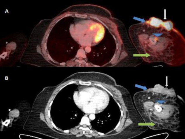
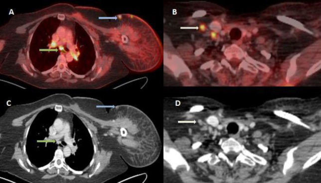
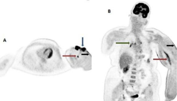
Objectives: Lymphangiosarcoma is a rare tumor that affects the upper limbs of patients who have undergone breast cancer surgery, including axillary dissection, followed by radiation therapy (RT) to the axilla and has a poor prognosis. 18F-FDG PET/CT may enable the earlier detection of malignant transformation in a setting of chronic lymphedema and help evaluate the extent and staging of the tumor, allowing earlier initiation of treatment options.
Case presentation: We herein report a case of cutaneous lymphangiosarcoma in a 47-year-old breast carcinoma patient, which occurred 9 years after initial surgery and radiation therapy. Distant metastases were detected on 18F-FDG PET/CT. The patient underwent fore-quarter amputation of the upper limb and concurrent chemo-radiation therapy. However, she succumbed to her disease after 3 cycles of chemotherapy.
Conclusions: 18F-FDG PET/CT scan helps in the early detection of malignant transformation and lymphangiosarcoma in a setting of chronic lymphedema in breast carcinoma patients following radiation therapy to the axilla. Furthermore, it helps determine the extent of regional spread and detect metastatic involvement, thus enabling better clinical management of these patients.

 求助内容:
求助内容: 应助结果提醒方式:


