Kelly N. Messingham, Paige T. Richards, Anthony Fleck, Radhika A. Patel, Marija Djurkovic, Jonah Elliff, Samuel Connell, Tyler P. Crowe, Juan Munoz Gonzalez, Francoise Gourronc, Jacob A. Dillard, Robert A. Davey, Aloysius Klingelhutz, Olena Shtanko, Wendy Maury
{"title":"多种细胞类型支持生产性感染和传染性埃博拉病毒向人体皮肤表面的动态易位","authors":"Kelly N. Messingham, Paige T. Richards, Anthony Fleck, Radhika A. Patel, Marija Djurkovic, Jonah Elliff, Samuel Connell, Tyler P. Crowe, Juan Munoz Gonzalez, Francoise Gourronc, Jacob A. Dillard, Robert A. Davey, Aloysius Klingelhutz, Olena Shtanko, Wendy Maury","doi":"10.1126/sciadv.adr6140","DOIUrl":null,"url":null,"abstract":"<div >Ebola virus (EBOV) causes severe human disease. During late infection, EBOV virions are on the skin’s surface; however, the permissive skin cell types and the route of virus translocation to the epidermal surface are unknown. We describe a human skin explant model and demonstrate that EBOV infection of human skin via basal media increases in a time-dependent and dose-dependent manner. In the dermis, cells of myeloid, endothelial, and fibroblast origin were EBOV antigen–positive whereas keratinocytes harbored virus in the epidermis. Infectious virus was detected on the apical epidermal surface within 3 days, indicating that virus propagates and traffics through the explants. Purified human fibroblasts and keratinocytes supported EBOV infection ex vivo and both cell types required the phosphatidylserine receptor, AXL, and the endosomal protein, NPC1, for virus entry. This platform identified susceptible cell types and demonstrated dynamic trafficking of EBOV virions. These findings may explain person-to-person transmission via skin contact.</div>","PeriodicalId":21609,"journal":{"name":"Science Advances","volume":"11 1","pages":""},"PeriodicalIF":12.5000,"publicationDate":"2025-01-01","publicationTypes":"Journal Article","fieldsOfStudy":null,"isOpenAccess":false,"openAccessPdf":"https://www.science.org/doi/reader/10.1126/sciadv.adr6140","citationCount":"0","resultStr":"{\"title\":\"Multiple cell types support productive infection and dynamic translocation of infectious Ebola virus to the surface of human skin\",\"authors\":\"Kelly N. Messingham, Paige T. Richards, Anthony Fleck, Radhika A. Patel, Marija Djurkovic, Jonah Elliff, Samuel Connell, Tyler P. Crowe, Juan Munoz Gonzalez, Francoise Gourronc, Jacob A. Dillard, Robert A. Davey, Aloysius Klingelhutz, Olena Shtanko, Wendy Maury\",\"doi\":\"10.1126/sciadv.adr6140\",\"DOIUrl\":null,\"url\":null,\"abstract\":\"<div >Ebola virus (EBOV) causes severe human disease. During late infection, EBOV virions are on the skin’s surface; however, the permissive skin cell types and the route of virus translocation to the epidermal surface are unknown. We describe a human skin explant model and demonstrate that EBOV infection of human skin via basal media increases in a time-dependent and dose-dependent manner. In the dermis, cells of myeloid, endothelial, and fibroblast origin were EBOV antigen–positive whereas keratinocytes harbored virus in the epidermis. Infectious virus was detected on the apical epidermal surface within 3 days, indicating that virus propagates and traffics through the explants. Purified human fibroblasts and keratinocytes supported EBOV infection ex vivo and both cell types required the phosphatidylserine receptor, AXL, and the endosomal protein, NPC1, for virus entry. This platform identified susceptible cell types and demonstrated dynamic trafficking of EBOV virions. These findings may explain person-to-person transmission via skin contact.</div>\",\"PeriodicalId\":21609,\"journal\":{\"name\":\"Science Advances\",\"volume\":\"11 1\",\"pages\":\"\"},\"PeriodicalIF\":12.5000,\"publicationDate\":\"2025-01-01\",\"publicationTypes\":\"Journal Article\",\"fieldsOfStudy\":null,\"isOpenAccess\":false,\"openAccessPdf\":\"https://www.science.org/doi/reader/10.1126/sciadv.adr6140\",\"citationCount\":\"0\",\"resultStr\":null,\"platform\":\"Semanticscholar\",\"paperid\":null,\"PeriodicalName\":\"Science Advances\",\"FirstCategoryId\":\"103\",\"ListUrlMain\":\"https://www.science.org/doi/10.1126/sciadv.adr6140\",\"RegionNum\":1,\"RegionCategory\":\"综合性期刊\",\"ArticlePicture\":[],\"TitleCN\":null,\"AbstractTextCN\":null,\"PMCID\":null,\"EPubDate\":\"\",\"PubModel\":\"\",\"JCR\":\"Q1\",\"JCRName\":\"MULTIDISCIPLINARY SCIENCES\",\"Score\":null,\"Total\":0}","platform":"Semanticscholar","paperid":null,"PeriodicalName":"Science Advances","FirstCategoryId":"103","ListUrlMain":"https://www.science.org/doi/10.1126/sciadv.adr6140","RegionNum":1,"RegionCategory":"综合性期刊","ArticlePicture":[],"TitleCN":null,"AbstractTextCN":null,"PMCID":null,"EPubDate":"","PubModel":"","JCR":"Q1","JCRName":"MULTIDISCIPLINARY SCIENCES","Score":null,"Total":0}
Multiple cell types support productive infection and dynamic translocation of infectious Ebola virus to the surface of human skin
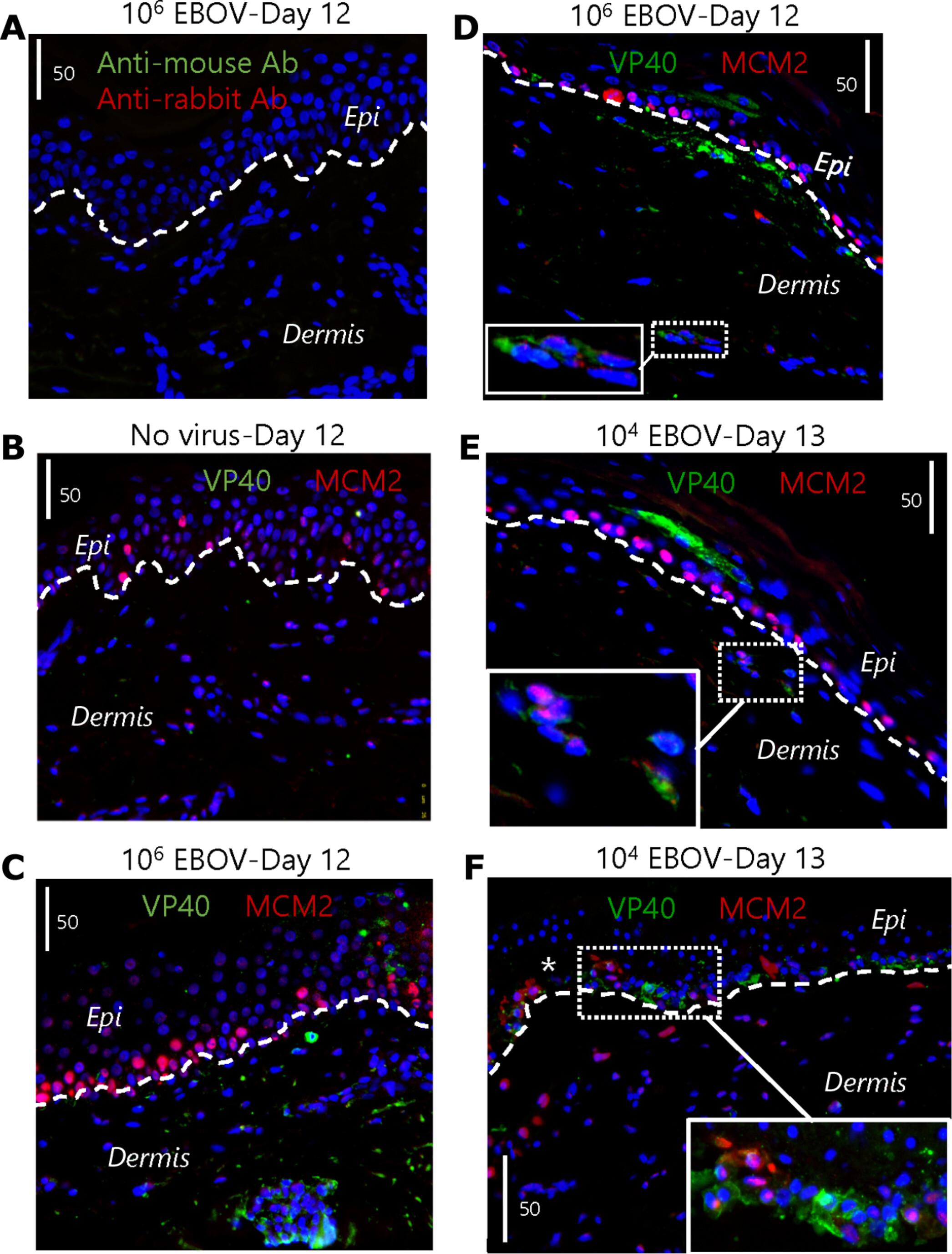
Ebola virus (EBOV) causes severe human disease. During late infection, EBOV virions are on the skin’s surface; however, the permissive skin cell types and the route of virus translocation to the epidermal surface are unknown. We describe a human skin explant model and demonstrate that EBOV infection of human skin via basal media increases in a time-dependent and dose-dependent manner. In the dermis, cells of myeloid, endothelial, and fibroblast origin were EBOV antigen–positive whereas keratinocytes harbored virus in the epidermis. Infectious virus was detected on the apical epidermal surface within 3 days, indicating that virus propagates and traffics through the explants. Purified human fibroblasts and keratinocytes supported EBOV infection ex vivo and both cell types required the phosphatidylserine receptor, AXL, and the endosomal protein, NPC1, for virus entry. This platform identified susceptible cell types and demonstrated dynamic trafficking of EBOV virions. These findings may explain person-to-person transmission via skin contact.
期刊介绍:
Science Advances, an open-access journal by AAAS, publishes impactful research in diverse scientific areas. It aims for fair, fast, and expert peer review, providing freely accessible research to readers. Led by distinguished scientists, the journal supports AAAS's mission by extending Science magazine's capacity to identify and promote significant advances. Evolving digital publishing technologies play a crucial role in advancing AAAS's global mission for science communication and benefitting humankind.