Tatjana Vilibic-Cavlek, Maja Bogdanic, Vladimir Savic, Zeljka Hruskar, Ljubo Barbic, Vladimir Stevanovic, Ljiljana Antolasic, Ljiljana Milasincic, Dario Sabadi, Gorana Miletic, Ivona Coric, Anna Mrzljak, Eddy Listes, Giovanni Savini
{"title":"西尼罗病毒感染的诊断:不同实验室方法的评价。","authors":"Tatjana Vilibic-Cavlek, Maja Bogdanic, Vladimir Savic, Zeljka Hruskar, Ljubo Barbic, Vladimir Stevanovic, Ljiljana Antolasic, Ljiljana Milasincic, Dario Sabadi, Gorana Miletic, Ivona Coric, Anna Mrzljak, Eddy Listes, Giovanni Savini","doi":"10.5501/wjv.v13.i4.95986","DOIUrl":null,"url":null,"abstract":"<p><strong>Background: </strong>The diagnosis of West Nile virus (WNV) is challenging due to short-term and low-level viremia, flavivirus cross-reactivity, and long immunoglobulin M (IgM) persistence.</p><p><strong>Aim: </strong>To evaluate different methods for WNV detection [reverse transcription-polymerase chain reaction (RT-PCR), IgM/IgG antibodies, IgG avidity] in serum, cerebrospinal fluid (CSF), and urine samples of patients with confirmed WNV infection.</p><p><strong>Methods: </strong>The study included patients with confirmed WNV neuroinvasive infection (<i>n</i> = 62), asymptomatic WNV seropositive individuals (<i>n</i> = 22), and individuals with false-positive WNV IgM antibodies (<i>n</i> = 30). WNV RNA was detected using RT-PCR. A commercial ELISA was used to detect WNV IgM/IgG antibodies with confirmation of cross-reactive samples using a virus neutralization test (VNT). IgG-positive samples were tested for IgG avidity.</p><p><strong>Results: </strong>The WNV-RNA detection rates were significantly higher in the urine (54.5%)/serum (46.4%) than in CSF (32.2%). According to the sampling time, the WNV-RNA detection rates in urine collected within 7 days/8-14/≥ 15 days were 29.4/66.6/62.5% (<i>P</i> = 0.042). However, these differences were not observed in the CSF. The median RT-PCR cycle threshold values were significantly lower in urine (32.5, IQR = 28-34) than in CSF (34.5, IQR = 33-36). The frequency of positive WNV IgM and IgG significantly differed according to the sampling time in serum but not in CSF. Positive IgM/IgG antibodies were detected in 84.3/9.3% of serum samples collected within 7 days, 100/71.1% of samples collected 8-14, and 100% samples collected after ≥ 15 days. Recent WNV infection was confirmed by low/borderline avidity index (AI) in 13.6% of asymptomatic individuals. A correlation between ELISA and AI was strong negative for IgM and strong positive for IgG. No significant correlation between ELISA IgG and VNT was found.</p><p><strong>Conclusion: </strong>The frequency of WNV RNA and antibody detection depends on the sampling time and type of clinical samples. IgG avidity could differentiate recent WNV infections from long-persisting IgM antibodies.</p>","PeriodicalId":61903,"journal":{"name":"世界病毒学杂志(英文版)","volume":"13 4","pages":"95986"},"PeriodicalIF":0.0000,"publicationDate":"2024-12-25","publicationTypes":"Journal Article","fieldsOfStudy":null,"isOpenAccess":false,"openAccessPdf":"https://www.ncbi.nlm.nih.gov/pmc/articles/PMC11551685/pdf/","citationCount":"0","resultStr":"{\"title\":\"Diagnosis of West Nile virus infections: Evaluation of different laboratory methods.\",\"authors\":\"Tatjana Vilibic-Cavlek, Maja Bogdanic, Vladimir Savic, Zeljka Hruskar, Ljubo Barbic, Vladimir Stevanovic, Ljiljana Antolasic, Ljiljana Milasincic, Dario Sabadi, Gorana Miletic, Ivona Coric, Anna Mrzljak, Eddy Listes, Giovanni Savini\",\"doi\":\"10.5501/wjv.v13.i4.95986\",\"DOIUrl\":null,\"url\":null,\"abstract\":\"<p><strong>Background: </strong>The diagnosis of West Nile virus (WNV) is challenging due to short-term and low-level viremia, flavivirus cross-reactivity, and long immunoglobulin M (IgM) persistence.</p><p><strong>Aim: </strong>To evaluate different methods for WNV detection [reverse transcription-polymerase chain reaction (RT-PCR), IgM/IgG antibodies, IgG avidity] in serum, cerebrospinal fluid (CSF), and urine samples of patients with confirmed WNV infection.</p><p><strong>Methods: </strong>The study included patients with confirmed WNV neuroinvasive infection (<i>n</i> = 62), asymptomatic WNV seropositive individuals (<i>n</i> = 22), and individuals with false-positive WNV IgM antibodies (<i>n</i> = 30). WNV RNA was detected using RT-PCR. A commercial ELISA was used to detect WNV IgM/IgG antibodies with confirmation of cross-reactive samples using a virus neutralization test (VNT). IgG-positive samples were tested for IgG avidity.</p><p><strong>Results: </strong>The WNV-RNA detection rates were significantly higher in the urine (54.5%)/serum (46.4%) than in CSF (32.2%). According to the sampling time, the WNV-RNA detection rates in urine collected within 7 days/8-14/≥ 15 days were 29.4/66.6/62.5% (<i>P</i> = 0.042). However, these differences were not observed in the CSF. The median RT-PCR cycle threshold values were significantly lower in urine (32.5, IQR = 28-34) than in CSF (34.5, IQR = 33-36). The frequency of positive WNV IgM and IgG significantly differed according to the sampling time in serum but not in CSF. Positive IgM/IgG antibodies were detected in 84.3/9.3% of serum samples collected within 7 days, 100/71.1% of samples collected 8-14, and 100% samples collected after ≥ 15 days. Recent WNV infection was confirmed by low/borderline avidity index (AI) in 13.6% of asymptomatic individuals. A correlation between ELISA and AI was strong negative for IgM and strong positive for IgG. No significant correlation between ELISA IgG and VNT was found.</p><p><strong>Conclusion: </strong>The frequency of WNV RNA and antibody detection depends on the sampling time and type of clinical samples. IgG avidity could differentiate recent WNV infections from long-persisting IgM antibodies.</p>\",\"PeriodicalId\":61903,\"journal\":{\"name\":\"世界病毒学杂志(英文版)\",\"volume\":\"13 4\",\"pages\":\"95986\"},\"PeriodicalIF\":0.0000,\"publicationDate\":\"2024-12-25\",\"publicationTypes\":\"Journal Article\",\"fieldsOfStudy\":null,\"isOpenAccess\":false,\"openAccessPdf\":\"https://www.ncbi.nlm.nih.gov/pmc/articles/PMC11551685/pdf/\",\"citationCount\":\"0\",\"resultStr\":null,\"platform\":\"Semanticscholar\",\"paperid\":null,\"PeriodicalName\":\"世界病毒学杂志(英文版)\",\"FirstCategoryId\":\"1089\",\"ListUrlMain\":\"https://doi.org/10.5501/wjv.v13.i4.95986\",\"RegionNum\":0,\"RegionCategory\":null,\"ArticlePicture\":[],\"TitleCN\":null,\"AbstractTextCN\":null,\"PMCID\":null,\"EPubDate\":\"\",\"PubModel\":\"\",\"JCR\":\"\",\"JCRName\":\"\",\"Score\":null,\"Total\":0}","platform":"Semanticscholar","paperid":null,"PeriodicalName":"世界病毒学杂志(英文版)","FirstCategoryId":"1089","ListUrlMain":"https://doi.org/10.5501/wjv.v13.i4.95986","RegionNum":0,"RegionCategory":null,"ArticlePicture":[],"TitleCN":null,"AbstractTextCN":null,"PMCID":null,"EPubDate":"","PubModel":"","JCR":"","JCRName":"","Score":null,"Total":0}
Diagnosis of West Nile virus infections: Evaluation of different laboratory methods.
Background: The diagnosis of West Nile virus (WNV) is challenging due to short-term and low-level viremia, flavivirus cross-reactivity, and long immunoglobulin M (IgM) persistence.
Aim: To evaluate different methods for WNV detection [reverse transcription-polymerase chain reaction (RT-PCR), IgM/IgG antibodies, IgG avidity] in serum, cerebrospinal fluid (CSF), and urine samples of patients with confirmed WNV infection.
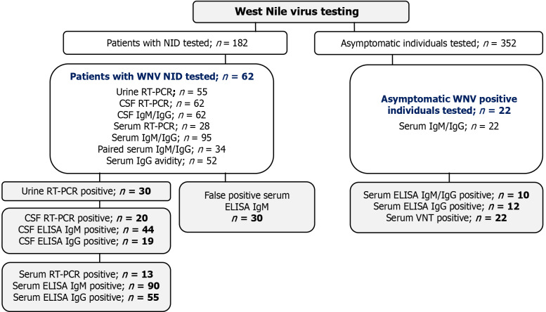
Methods: The study included patients with confirmed WNV neuroinvasive infection (n = 62), asymptomatic WNV seropositive individuals (n = 22), and individuals with false-positive WNV IgM antibodies (n = 30). WNV RNA was detected using RT-PCR. A commercial ELISA was used to detect WNV IgM/IgG antibodies with confirmation of cross-reactive samples using a virus neutralization test (VNT). IgG-positive samples were tested for IgG avidity.
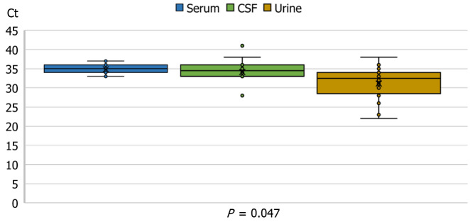
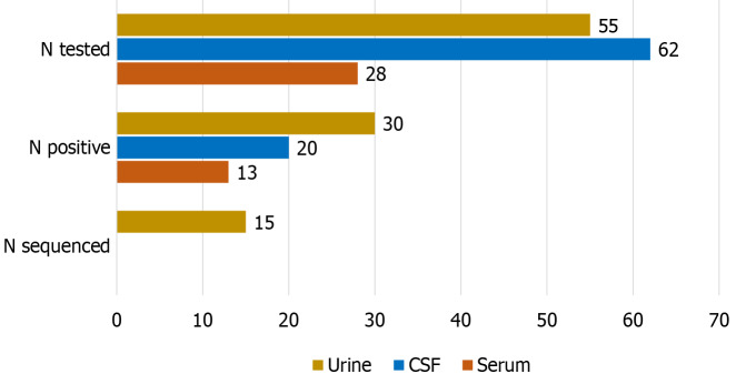
Results: The WNV-RNA detection rates were significantly higher in the urine (54.5%)/serum (46.4%) than in CSF (32.2%). According to the sampling time, the WNV-RNA detection rates in urine collected within 7 days/8-14/≥ 15 days were 29.4/66.6/62.5% (P = 0.042). However, these differences were not observed in the CSF. The median RT-PCR cycle threshold values were significantly lower in urine (32.5, IQR = 28-34) than in CSF (34.5, IQR = 33-36). The frequency of positive WNV IgM and IgG significantly differed according to the sampling time in serum but not in CSF. Positive IgM/IgG antibodies were detected in 84.3/9.3% of serum samples collected within 7 days, 100/71.1% of samples collected 8-14, and 100% samples collected after ≥ 15 days. Recent WNV infection was confirmed by low/borderline avidity index (AI) in 13.6% of asymptomatic individuals. A correlation between ELISA and AI was strong negative for IgM and strong positive for IgG. No significant correlation between ELISA IgG and VNT was found.
Conclusion: The frequency of WNV RNA and antibody detection depends on the sampling time and type of clinical samples. IgG avidity could differentiate recent WNV infections from long-persisting IgM antibodies.

 求助内容:
求助内容: 应助结果提醒方式:


