{"title":"单纯心电图参数与阵发性心房颤动的关系。","authors":"Mohammad Assadian Rad, Hanie Shadrou, Sajad Kazemalilou, Habib Eslami Kenarsari, Mahboobeh Gholipour","doi":"10.48305/arya.2024.41690.2900","DOIUrl":null,"url":null,"abstract":"<p><strong>Background: </strong>Atrial fibrillation (AF) is a prevalent arrhythmia, and predicting its occurrence plays a crucial role in reducing its complications. This study aimed to investigate the relation between simple P wave parameters and paroxysmal AF (pAF).</p><p><strong>Methods: </strong>In this case-control study, demographic and laboratory data were gathered by a checklist. P wave parameters were measured in electrocardiography (ECG). The relationship between these parameters and AF in groups was analyzed.</p><p><strong>Results: </strong>Eighty individuals were included (40 patients with pAF (57.5% female, mean age = 64.9 ± 2.04) and 40 individuals without AF (57.5% female, mean age = 60.3 ± 2.01)). The P wave peak time (PWPT) in leads D2 (p = 0.003) and V1 (p = 0.001) were longer in the case group. In addition, the prolongation of the PR interval (PR) in lead D2, P wave duration (PWD) in lead D2, and P terminal force (PTF) in V1 were associated with an increase in the occurrence of pAF. Adjusted regression analysis showed that two variables, PWPT in V1 (OR, 95% CI: 1.04 (1.01-1.07), p = 0.005) and PWD in D2 (OR, 95% CI: 1.03 (1.00-1.05), p = 0.018), were predictors for AF.</p><p><strong>Conclusion: </strong>Our results underscore the potential utility of simple ECG parameters, especially PWD in lead D2 and PWPT in V1, in predicting and assessing the risk of pAF. These findings provide valuable insights for clinical practice and risk stratification in patients without structural cardiac disease. Additionally, these findings may potentially contribute to the prevention of complications and injuries associated with pAF.</p>","PeriodicalId":46477,"journal":{"name":"ARYA Atherosclerosis","volume":"20 5","pages":"6-14"},"PeriodicalIF":0.6000,"publicationDate":"2024-01-01","publicationTypes":"Journal Article","fieldsOfStudy":null,"isOpenAccess":false,"openAccessPdf":"https://www.ncbi.nlm.nih.gov/pmc/articles/PMC11663446/pdf/","citationCount":"0","resultStr":"{\"title\":\"Relationship between simple electrocardiographic parameter and paroxysmal atrial fibrillation.\",\"authors\":\"Mohammad Assadian Rad, Hanie Shadrou, Sajad Kazemalilou, Habib Eslami Kenarsari, Mahboobeh Gholipour\",\"doi\":\"10.48305/arya.2024.41690.2900\",\"DOIUrl\":null,\"url\":null,\"abstract\":\"<p><strong>Background: </strong>Atrial fibrillation (AF) is a prevalent arrhythmia, and predicting its occurrence plays a crucial role in reducing its complications. This study aimed to investigate the relation between simple P wave parameters and paroxysmal AF (pAF).</p><p><strong>Methods: </strong>In this case-control study, demographic and laboratory data were gathered by a checklist. P wave parameters were measured in electrocardiography (ECG). The relationship between these parameters and AF in groups was analyzed.</p><p><strong>Results: </strong>Eighty individuals were included (40 patients with pAF (57.5% female, mean age = 64.9 ± 2.04) and 40 individuals without AF (57.5% female, mean age = 60.3 ± 2.01)). The P wave peak time (PWPT) in leads D2 (p = 0.003) and V1 (p = 0.001) were longer in the case group. In addition, the prolongation of the PR interval (PR) in lead D2, P wave duration (PWD) in lead D2, and P terminal force (PTF) in V1 were associated with an increase in the occurrence of pAF. Adjusted regression analysis showed that two variables, PWPT in V1 (OR, 95% CI: 1.04 (1.01-1.07), p = 0.005) and PWD in D2 (OR, 95% CI: 1.03 (1.00-1.05), p = 0.018), were predictors for AF.</p><p><strong>Conclusion: </strong>Our results underscore the potential utility of simple ECG parameters, especially PWD in lead D2 and PWPT in V1, in predicting and assessing the risk of pAF. These findings provide valuable insights for clinical practice and risk stratification in patients without structural cardiac disease. Additionally, these findings may potentially contribute to the prevention of complications and injuries associated with pAF.</p>\",\"PeriodicalId\":46477,\"journal\":{\"name\":\"ARYA Atherosclerosis\",\"volume\":\"20 5\",\"pages\":\"6-14\"},\"PeriodicalIF\":0.6000,\"publicationDate\":\"2024-01-01\",\"publicationTypes\":\"Journal Article\",\"fieldsOfStudy\":null,\"isOpenAccess\":false,\"openAccessPdf\":\"https://www.ncbi.nlm.nih.gov/pmc/articles/PMC11663446/pdf/\",\"citationCount\":\"0\",\"resultStr\":null,\"platform\":\"Semanticscholar\",\"paperid\":null,\"PeriodicalName\":\"ARYA Atherosclerosis\",\"FirstCategoryId\":\"1085\",\"ListUrlMain\":\"https://doi.org/10.48305/arya.2024.41690.2900\",\"RegionNum\":0,\"RegionCategory\":null,\"ArticlePicture\":[],\"TitleCN\":null,\"AbstractTextCN\":null,\"PMCID\":null,\"EPubDate\":\"\",\"PubModel\":\"\",\"JCR\":\"Q4\",\"JCRName\":\"CARDIAC & CARDIOVASCULAR SYSTEMS\",\"Score\":null,\"Total\":0}","platform":"Semanticscholar","paperid":null,"PeriodicalName":"ARYA Atherosclerosis","FirstCategoryId":"1085","ListUrlMain":"https://doi.org/10.48305/arya.2024.41690.2900","RegionNum":0,"RegionCategory":null,"ArticlePicture":[],"TitleCN":null,"AbstractTextCN":null,"PMCID":null,"EPubDate":"","PubModel":"","JCR":"Q4","JCRName":"CARDIAC & CARDIOVASCULAR SYSTEMS","Score":null,"Total":0}
Relationship between simple electrocardiographic parameter and paroxysmal atrial fibrillation.
Background: Atrial fibrillation (AF) is a prevalent arrhythmia, and predicting its occurrence plays a crucial role in reducing its complications. This study aimed to investigate the relation between simple P wave parameters and paroxysmal AF (pAF).
Methods: In this case-control study, demographic and laboratory data were gathered by a checklist. P wave parameters were measured in electrocardiography (ECG). The relationship between these parameters and AF in groups was analyzed.
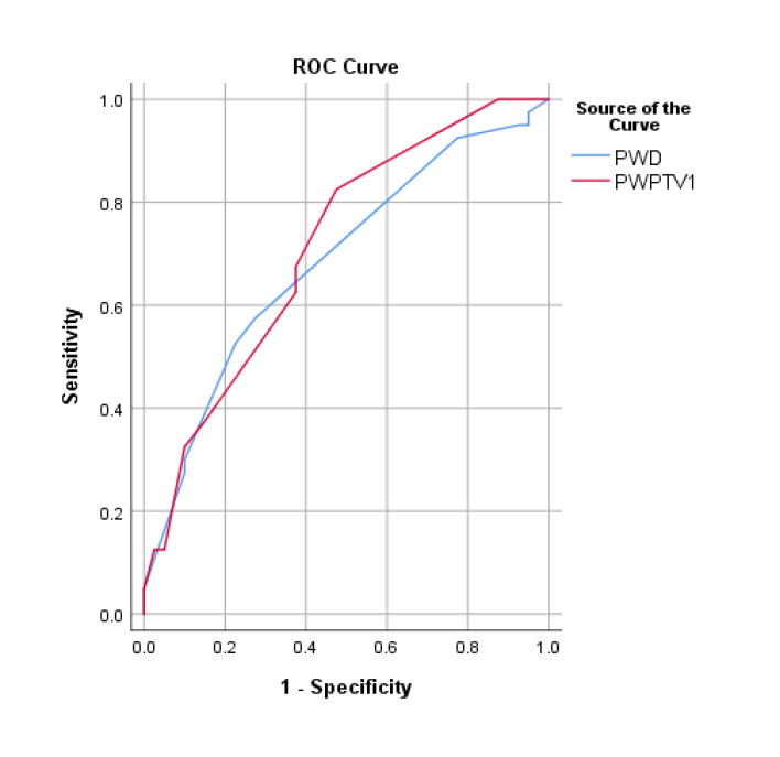
Results: Eighty individuals were included (40 patients with pAF (57.5% female, mean age = 64.9 ± 2.04) and 40 individuals without AF (57.5% female, mean age = 60.3 ± 2.01)). The P wave peak time (PWPT) in leads D2 (p = 0.003) and V1 (p = 0.001) were longer in the case group. In addition, the prolongation of the PR interval (PR) in lead D2, P wave duration (PWD) in lead D2, and P terminal force (PTF) in V1 were associated with an increase in the occurrence of pAF. Adjusted regression analysis showed that two variables, PWPT in V1 (OR, 95% CI: 1.04 (1.01-1.07), p = 0.005) and PWD in D2 (OR, 95% CI: 1.03 (1.00-1.05), p = 0.018), were predictors for AF.
Conclusion: Our results underscore the potential utility of simple ECG parameters, especially PWD in lead D2 and PWPT in V1, in predicting and assessing the risk of pAF. These findings provide valuable insights for clinical practice and risk stratification in patients without structural cardiac disease. Additionally, these findings may potentially contribute to the prevention of complications and injuries associated with pAF.

 求助内容:
求助内容: 应助结果提醒方式:


