通过三维时空统计形状模型探索健康骨骼生长中的形状变化:范围综述。
IF 2.6
Q3 ENDOCRINOLOGY & METABOLISM
引用次数: 0
摘要
目的:分析人群骨形态变化趋势,为研究生长过程提供有价值的依据。本文综述了最新的时空统计形状建模技术,强调了它们在健康生长过程中三维骨骼结构的应用。方法:我们使用儿童三维健康骨骼模型时空数据集在PubMed和Scopus上检索统计形状建模的文章。提取了数据集特征和形状模型开发、分析和潜在临床应用的细节。结果:14项研究被发现符合条件,模拟了一个或多个下肢骨骼、下颌骨、颅骨和椎骨。主要是利用主成分分析法对点分布模型建立统计形状模型。基于形状模态分析形状变化,将特定形状变化表示为整体变化的一部分。非比例模型产生的统计形状模型比比例模型更紧凑。后者代表了更微妙的形状变化,因为骨骼模型之间没有大小差异。四项研究报告了第一种形状模式与年龄之间的显著相关性,表明这种形状变化与生长之间的关系。三项研究利用统计形状建模的预测特征重构三维模型。测量预测和实际解剖结构之间的差异导致均方根误差小于3毫米。结论:时空统计形状建模提供了对生长过程中形状变化模式的洞察。这样的模型可以用来寻找预测因素,如年龄或性别,并利用这些特征来预测某人的骨骼几何形状。本文章由计算机程序翻译,如有差异,请以英文原文为准。
Exploring shape changes in healthy bone growth through 3D spatiotemporal statistical shape models: A scoping review
Objective
Analyzing population trends of bone shape variation can provide valuable insights into growth processes. This review aims to overview state-of-the-art spatiotemporal statistical shape modeling techniques, emphasizing their application to 3D skeletal structures during healthy growth.
Methods
We searched PubMed and Scopus for articles on statistical shape modeling using a pediatric spatiotemporal dataset of 3D healthy bone models. Dataset characteristics and details on the shape models' development, analyses, and potential clinical use were extracted.
Results
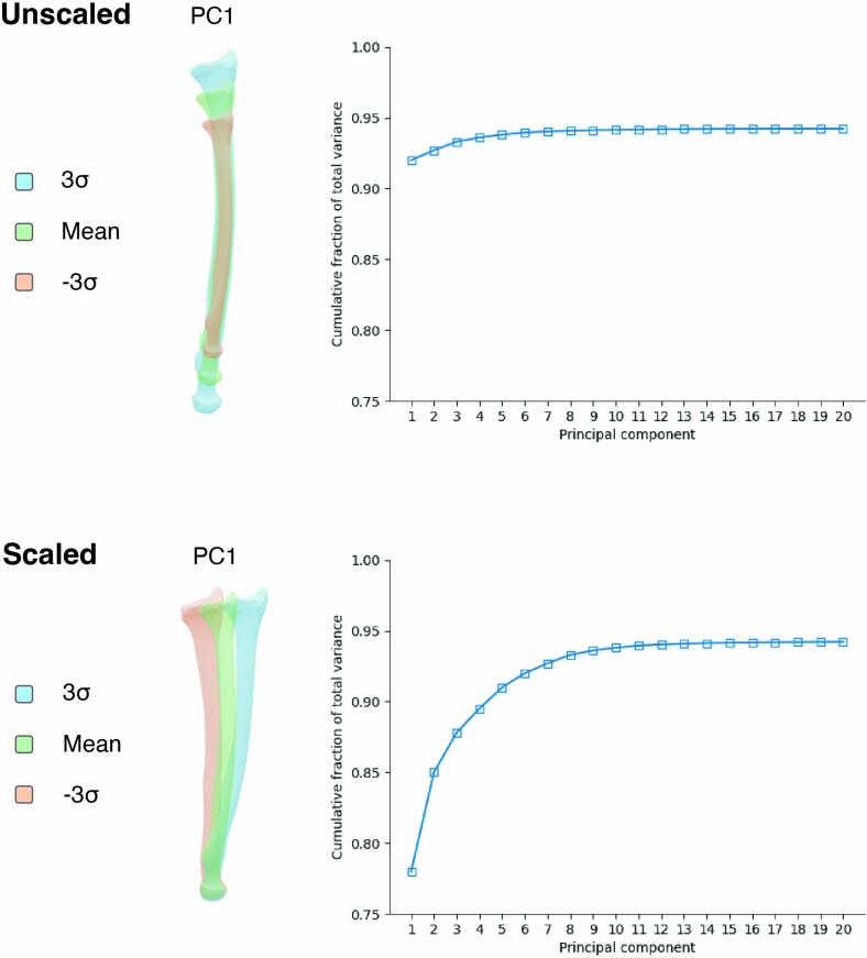
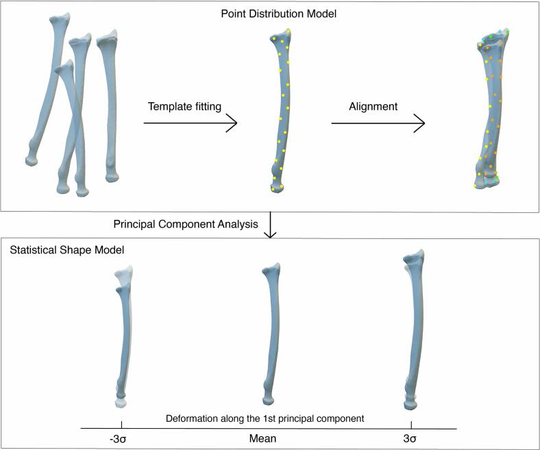
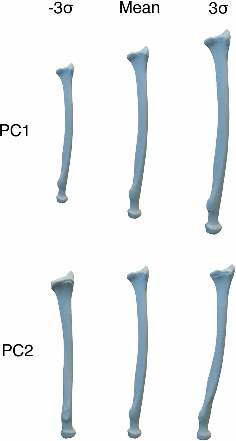
Fourteen studies were found eligible, modeling one or multiple lower limb bones, the mandible, the skull, and vertebrae. The majority applied Principal Component Analysis on point distribution models to create a statistical shape model. Shape variation was analyzed based on shape modes, representing a specific shape change as a part of the overall variance. Unscaled models resulted in a more compact statistical shape model than scaled models. The latter represented more subtle shape variations due to the absence of size differences between the bone models. Four studies reported a significant correlation between the first shape mode and age, indicating a relationship between that type of shape variation and growth. Three studies reconstructed 3D models using prediction features of statistical shape modeling. Measuring difference between predicted and actual anatomy resulted in Root Mean Squared Errors below 3 mm.
Conclusion
Spatiotemporal statistical shape modeling provides insight into modes of shape variation during growth. Such a model can be used to find predictive factors, like age or sex, and deploy these characteristics to predict someone's bone geometry.
求助全文
通过发布文献求助,成功后即可免费获取论文全文。
去求助
来源期刊
Bone Reports
Medicine-Orthopedics and Sports Medicine
CiteScore
4.30
自引率
4.00%
发文量
444
审稿时长
57 days
期刊介绍:
Bone Reports is an interdisciplinary forum for the rapid publication of Original Research Articles and Case Reports across basic, translational and clinical aspects of bone and mineral metabolism. The journal publishes papers that are scientifically sound, with the peer review process focused principally on verifying sound methodologies, and correct data analysis and interpretation. We welcome studies either replicating or failing to replicate a previous study, and null findings. We fulfil a critical and current need to enhance research by publishing reproducibility studies and null findings.
 求助内容:
求助内容: 应助结果提醒方式:


