Meagan Wu, Connor S Wagner, Dillan F Villavisanis, Jinggang J Ng, Benjamin B Massenburg, Dominic J Romeo, Gregory G Heuer, Scott P Bartlett, Jordan W Swanson, Jesse A Taylor
{"title":"内窥镜辅助与开放式额眶牵张治疗单冠状颅缝闭锁:形态学和技术考虑。","authors":"Meagan Wu, Connor S Wagner, Dillan F Villavisanis, Jinggang J Ng, Benjamin B Massenburg, Dominic J Romeo, Gregory G Heuer, Scott P Bartlett, Jordan W Swanson, Jesse A Taylor","doi":"10.1007/s00381-024-06662-8","DOIUrl":null,"url":null,"abstract":"<p><strong>Introduction: </strong>In an effort to maximize benefit and minimize morbidity when performing fronto-orbital distraction osteogenesis (FODO) for unilateral coronal synostosis (UCS), we have transitioned to an endoscopic-assisted approach (\"endo-FODO\"). This study compares photogrammetric outcomes of patients who underwent FODO via an endoscopic-assisted versus open approach.</p><p><strong>Methods: </strong>We retrospectively reviewed patients treated for UCS from 2013 to 2023. Photogrammetric outcomes at one to three years postoperatively were compared between patients who underwent endo-FODO and age- and sex-matched controls who underwent open FODO. Differences between pre- and postoperative periorbital symmetry ratios, canthal tilt symmetry, and orbital dystopia angle (ODA) were calculated.</p><p><strong>Results: </strong>Twenty patients (ten per group) underwent surgery at a mean age of 6.1 ± 1.8 and 5.4 ± 1.1 months (p = 0.426) and were photographed at 1.6 ± 0.9 and 1.8 ± 0.9 years (p = 0.597) postoperatively in the endo-FODO and open FODO groups, respectively. Patients who underwent endo-FODO demonstrated significant improvements in margin-reflex distance 1 (MRD1) symmetry ratio (p = 0.004), palpebral height symmetry ratio (p = 0.004), canthal tilt symmetry (p = 0.020), and ODA (p = 0.009). Patients who underwent open FODO likewise demonstrated significant improvements in MRD1 symmetry ratio (p = 0.004), palpebral height symmetry ratio (p = 0.033), and ODA (p = 0.004). All postoperative measurements as well as degrees of improvement were similar between groups (p > 0.05).</p><p><strong>Conclusions: </strong>Endo- and open FODO were associated with significant and comparable improvements in soft tissue periorbital symmetry and orbital dystopia at nearly two years postoperatively. While continued follow-up until cranial maturity is needed to assess the durability of aesthetic results, these data support a minimally invasive, endoscopic alternative to fronto-orbital distraction.</p>","PeriodicalId":9970,"journal":{"name":"Child's Nervous System","volume":"41 1","pages":"59"},"PeriodicalIF":1.2000,"publicationDate":"2024-12-18","publicationTypes":"Journal Article","fieldsOfStudy":null,"isOpenAccess":false,"openAccessPdf":"https://www.ncbi.nlm.nih.gov/pmc/articles/PMC11655604/pdf/","citationCount":"0","resultStr":"{\"title\":\"Endoscopic-assisted versus open fronto-orbital distraction for unicoronal craniosynostosis: morphometric and technique considerations.\",\"authors\":\"Meagan Wu, Connor S Wagner, Dillan F Villavisanis, Jinggang J Ng, Benjamin B Massenburg, Dominic J Romeo, Gregory G Heuer, Scott P Bartlett, Jordan W Swanson, Jesse A Taylor\",\"doi\":\"10.1007/s00381-024-06662-8\",\"DOIUrl\":null,\"url\":null,\"abstract\":\"<p><strong>Introduction: </strong>In an effort to maximize benefit and minimize morbidity when performing fronto-orbital distraction osteogenesis (FODO) for unilateral coronal synostosis (UCS), we have transitioned to an endoscopic-assisted approach (\\\"endo-FODO\\\"). This study compares photogrammetric outcomes of patients who underwent FODO via an endoscopic-assisted versus open approach.</p><p><strong>Methods: </strong>We retrospectively reviewed patients treated for UCS from 2013 to 2023. Photogrammetric outcomes at one to three years postoperatively were compared between patients who underwent endo-FODO and age- and sex-matched controls who underwent open FODO. Differences between pre- and postoperative periorbital symmetry ratios, canthal tilt symmetry, and orbital dystopia angle (ODA) were calculated.</p><p><strong>Results: </strong>Twenty patients (ten per group) underwent surgery at a mean age of 6.1 ± 1.8 and 5.4 ± 1.1 months (p = 0.426) and were photographed at 1.6 ± 0.9 and 1.8 ± 0.9 years (p = 0.597) postoperatively in the endo-FODO and open FODO groups, respectively. Patients who underwent endo-FODO demonstrated significant improvements in margin-reflex distance 1 (MRD1) symmetry ratio (p = 0.004), palpebral height symmetry ratio (p = 0.004), canthal tilt symmetry (p = 0.020), and ODA (p = 0.009). Patients who underwent open FODO likewise demonstrated significant improvements in MRD1 symmetry ratio (p = 0.004), palpebral height symmetry ratio (p = 0.033), and ODA (p = 0.004). All postoperative measurements as well as degrees of improvement were similar between groups (p > 0.05).</p><p><strong>Conclusions: </strong>Endo- and open FODO were associated with significant and comparable improvements in soft tissue periorbital symmetry and orbital dystopia at nearly two years postoperatively. While continued follow-up until cranial maturity is needed to assess the durability of aesthetic results, these data support a minimally invasive, endoscopic alternative to fronto-orbital distraction.</p>\",\"PeriodicalId\":9970,\"journal\":{\"name\":\"Child's Nervous System\",\"volume\":\"41 1\",\"pages\":\"59\"},\"PeriodicalIF\":1.2000,\"publicationDate\":\"2024-12-18\",\"publicationTypes\":\"Journal Article\",\"fieldsOfStudy\":null,\"isOpenAccess\":false,\"openAccessPdf\":\"https://www.ncbi.nlm.nih.gov/pmc/articles/PMC11655604/pdf/\",\"citationCount\":\"0\",\"resultStr\":null,\"platform\":\"Semanticscholar\",\"paperid\":null,\"PeriodicalName\":\"Child's Nervous System\",\"FirstCategoryId\":\"3\",\"ListUrlMain\":\"https://doi.org/10.1007/s00381-024-06662-8\",\"RegionNum\":4,\"RegionCategory\":\"医学\",\"ArticlePicture\":[],\"TitleCN\":null,\"AbstractTextCN\":null,\"PMCID\":null,\"EPubDate\":\"\",\"PubModel\":\"\",\"JCR\":\"Q4\",\"JCRName\":\"CLINICAL NEUROLOGY\",\"Score\":null,\"Total\":0}","platform":"Semanticscholar","paperid":null,"PeriodicalName":"Child's Nervous System","FirstCategoryId":"3","ListUrlMain":"https://doi.org/10.1007/s00381-024-06662-8","RegionNum":4,"RegionCategory":"医学","ArticlePicture":[],"TitleCN":null,"AbstractTextCN":null,"PMCID":null,"EPubDate":"","PubModel":"","JCR":"Q4","JCRName":"CLINICAL NEUROLOGY","Score":null,"Total":0}
Endoscopic-assisted versus open fronto-orbital distraction for unicoronal craniosynostosis: morphometric and technique considerations.
Introduction: In an effort to maximize benefit and minimize morbidity when performing fronto-orbital distraction osteogenesis (FODO) for unilateral coronal synostosis (UCS), we have transitioned to an endoscopic-assisted approach ("endo-FODO"). This study compares photogrammetric outcomes of patients who underwent FODO via an endoscopic-assisted versus open approach.
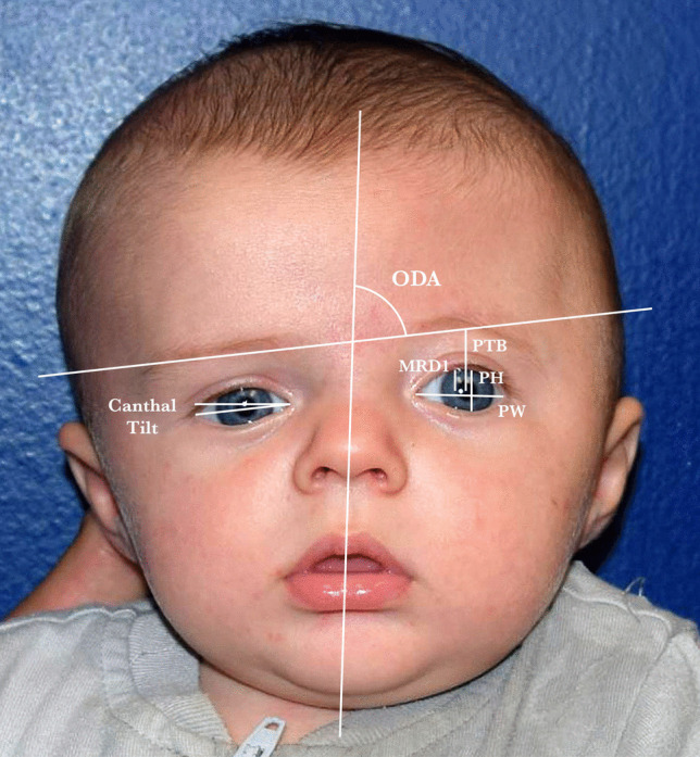
Methods: We retrospectively reviewed patients treated for UCS from 2013 to 2023. Photogrammetric outcomes at one to three years postoperatively were compared between patients who underwent endo-FODO and age- and sex-matched controls who underwent open FODO. Differences between pre- and postoperative periorbital symmetry ratios, canthal tilt symmetry, and orbital dystopia angle (ODA) were calculated.
Results: Twenty patients (ten per group) underwent surgery at a mean age of 6.1 ± 1.8 and 5.4 ± 1.1 months (p = 0.426) and were photographed at 1.6 ± 0.9 and 1.8 ± 0.9 years (p = 0.597) postoperatively in the endo-FODO and open FODO groups, respectively. Patients who underwent endo-FODO demonstrated significant improvements in margin-reflex distance 1 (MRD1) symmetry ratio (p = 0.004), palpebral height symmetry ratio (p = 0.004), canthal tilt symmetry (p = 0.020), and ODA (p = 0.009). Patients who underwent open FODO likewise demonstrated significant improvements in MRD1 symmetry ratio (p = 0.004), palpebral height symmetry ratio (p = 0.033), and ODA (p = 0.004). All postoperative measurements as well as degrees of improvement were similar between groups (p > 0.05).
Conclusions: Endo- and open FODO were associated with significant and comparable improvements in soft tissue periorbital symmetry and orbital dystopia at nearly two years postoperatively. While continued follow-up until cranial maturity is needed to assess the durability of aesthetic results, these data support a minimally invasive, endoscopic alternative to fronto-orbital distraction.
期刊介绍:
The journal has been expanded to encompass all aspects of pediatric neurosciences concerning the developmental and acquired abnormalities of the nervous system and its coverings, functional disorders, epilepsy, spasticity, basic and clinical neuro-oncology, rehabilitation and trauma. Global pediatric neurosurgery is an additional field of interest that will be considered for publication in the journal.
 求助内容:
求助内容: 应助结果提醒方式:


