John F Griffin, William S Stevenson, Nathan C Nelson, Annie V Chen, Silke Hecht, Brian F Porter, C Elizabeth Boudreau, Swan Specchi, Marco Bernardini, Wilfried Mai
{"title":"六只患有脑和脊髓上皮瘤的狗的核磁共振成像结果。","authors":"John F Griffin, William S Stevenson, Nathan C Nelson, Annie V Chen, Silke Hecht, Brian F Porter, C Elizabeth Boudreau, Swan Specchi, Marco Bernardini, Wilfried Mai","doi":"10.1111/vru.13477","DOIUrl":null,"url":null,"abstract":"<p><p>There are few published descriptions of the MRI appearance of canine intracranial or spinal cord ependymoma. In this multicenter, retrospective, secondary analysis, case series study, three veterinary radiologists independently reviewed and recorded imaging characteristics of MRI studies in six dogs with histopathologically confirmed ependymoma (three intracranial and three spinal cord cases). A consensus was reached when there was disagreement on specific features. All intracranial ependymomas had forebrain location, heterogeneous signal intensity in T1-weighted (T1W) and T2-weighted (T2W) images, heterogeneous contrast enhancement, and hyperintensity in T2W images. Two ependymomas had an intraventricular location; one was intra-axial. Other imaging features included intralesional cyst-like structures, intralesional hemorrhage, and perilesional edema. Dogs with spinal cord ependymoma had intramedullary lesions located in the cervical or thoracic spinal cord. Spinal cord ependymomas were isointense and homogeneous in T1W images and hyperintense in T2W images. Lesion location relative to the central canal of the spinal cord was variable. All three spinal cord ependymomas had perilesional T2W hyperintensity and moderate, heterogeneous contrast enhancement. None of the spinal cord ependymomas had intralesional cyst-like structures. One spinal cord ependymoma had evidence of drop metastases (diffuse, leptomeningeal). MRI features of canine ependymomas overlap with those of other diseases of the brain and spinal cord. Ependymoma should be considered a differential diagnosis for dogs with intraventricular, intra-axial forebrain, or intramedullary spinal cord masses.</p>","PeriodicalId":23581,"journal":{"name":"Veterinary Radiology & Ultrasound","volume":"66 1","pages":"e13477"},"PeriodicalIF":1.5000,"publicationDate":"2025-01-01","publicationTypes":"Journal Article","fieldsOfStudy":null,"isOpenAccess":false,"openAccessPdf":"https://www.ncbi.nlm.nih.gov/pmc/articles/PMC11649882/pdf/","citationCount":"0","resultStr":"{\"title\":\"MRI findings in six dogs with ependymoma of the brain and spinal cord.\",\"authors\":\"John F Griffin, William S Stevenson, Nathan C Nelson, Annie V Chen, Silke Hecht, Brian F Porter, C Elizabeth Boudreau, Swan Specchi, Marco Bernardini, Wilfried Mai\",\"doi\":\"10.1111/vru.13477\",\"DOIUrl\":null,\"url\":null,\"abstract\":\"<p><p>There are few published descriptions of the MRI appearance of canine intracranial or spinal cord ependymoma. In this multicenter, retrospective, secondary analysis, case series study, three veterinary radiologists independently reviewed and recorded imaging characteristics of MRI studies in six dogs with histopathologically confirmed ependymoma (three intracranial and three spinal cord cases). A consensus was reached when there was disagreement on specific features. All intracranial ependymomas had forebrain location, heterogeneous signal intensity in T1-weighted (T1W) and T2-weighted (T2W) images, heterogeneous contrast enhancement, and hyperintensity in T2W images. Two ependymomas had an intraventricular location; one was intra-axial. Other imaging features included intralesional cyst-like structures, intralesional hemorrhage, and perilesional edema. Dogs with spinal cord ependymoma had intramedullary lesions located in the cervical or thoracic spinal cord. Spinal cord ependymomas were isointense and homogeneous in T1W images and hyperintense in T2W images. Lesion location relative to the central canal of the spinal cord was variable. All three spinal cord ependymomas had perilesional T2W hyperintensity and moderate, heterogeneous contrast enhancement. None of the spinal cord ependymomas had intralesional cyst-like structures. One spinal cord ependymoma had evidence of drop metastases (diffuse, leptomeningeal). MRI features of canine ependymomas overlap with those of other diseases of the brain and spinal cord. Ependymoma should be considered a differential diagnosis for dogs with intraventricular, intra-axial forebrain, or intramedullary spinal cord masses.</p>\",\"PeriodicalId\":23581,\"journal\":{\"name\":\"Veterinary Radiology & Ultrasound\",\"volume\":\"66 1\",\"pages\":\"e13477\"},\"PeriodicalIF\":1.5000,\"publicationDate\":\"2025-01-01\",\"publicationTypes\":\"Journal Article\",\"fieldsOfStudy\":null,\"isOpenAccess\":false,\"openAccessPdf\":\"https://www.ncbi.nlm.nih.gov/pmc/articles/PMC11649882/pdf/\",\"citationCount\":\"0\",\"resultStr\":null,\"platform\":\"Semanticscholar\",\"paperid\":null,\"PeriodicalName\":\"Veterinary Radiology & Ultrasound\",\"FirstCategoryId\":\"97\",\"ListUrlMain\":\"https://doi.org/10.1111/vru.13477\",\"RegionNum\":2,\"RegionCategory\":\"农林科学\",\"ArticlePicture\":[],\"TitleCN\":null,\"AbstractTextCN\":null,\"PMCID\":null,\"EPubDate\":\"\",\"PubModel\":\"\",\"JCR\":\"Q2\",\"JCRName\":\"VETERINARY SCIENCES\",\"Score\":null,\"Total\":0}","platform":"Semanticscholar","paperid":null,"PeriodicalName":"Veterinary Radiology & Ultrasound","FirstCategoryId":"97","ListUrlMain":"https://doi.org/10.1111/vru.13477","RegionNum":2,"RegionCategory":"农林科学","ArticlePicture":[],"TitleCN":null,"AbstractTextCN":null,"PMCID":null,"EPubDate":"","PubModel":"","JCR":"Q2","JCRName":"VETERINARY SCIENCES","Score":null,"Total":0}
MRI findings in six dogs with ependymoma of the brain and spinal cord.
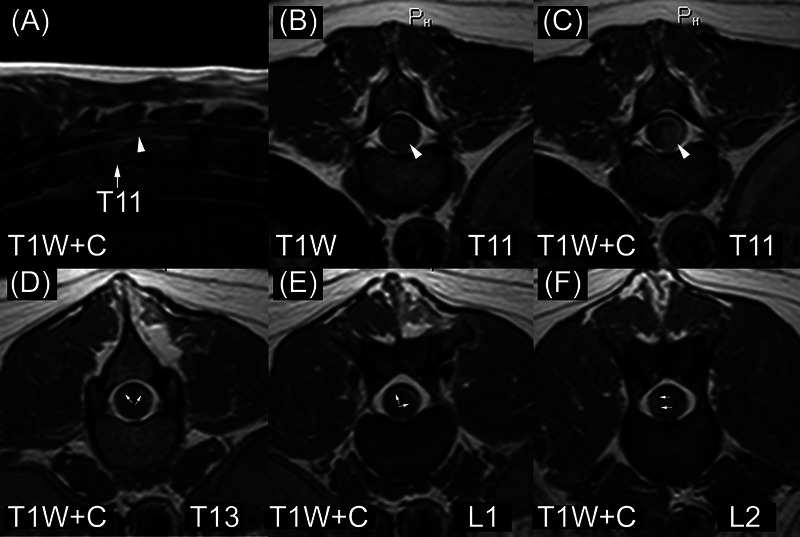
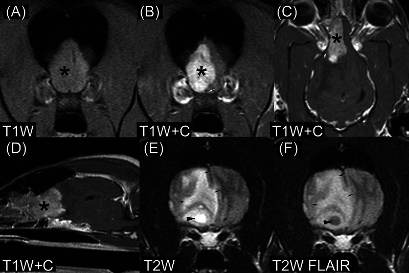
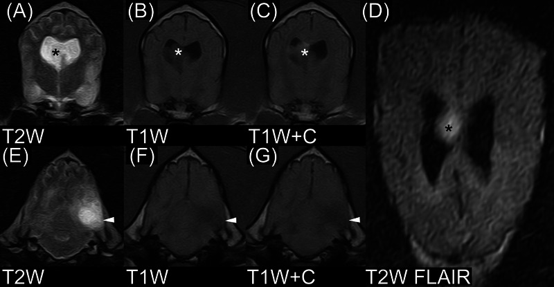
There are few published descriptions of the MRI appearance of canine intracranial or spinal cord ependymoma. In this multicenter, retrospective, secondary analysis, case series study, three veterinary radiologists independently reviewed and recorded imaging characteristics of MRI studies in six dogs with histopathologically confirmed ependymoma (three intracranial and three spinal cord cases). A consensus was reached when there was disagreement on specific features. All intracranial ependymomas had forebrain location, heterogeneous signal intensity in T1-weighted (T1W) and T2-weighted (T2W) images, heterogeneous contrast enhancement, and hyperintensity in T2W images. Two ependymomas had an intraventricular location; one was intra-axial. Other imaging features included intralesional cyst-like structures, intralesional hemorrhage, and perilesional edema. Dogs with spinal cord ependymoma had intramedullary lesions located in the cervical or thoracic spinal cord. Spinal cord ependymomas were isointense and homogeneous in T1W images and hyperintense in T2W images. Lesion location relative to the central canal of the spinal cord was variable. All three spinal cord ependymomas had perilesional T2W hyperintensity and moderate, heterogeneous contrast enhancement. None of the spinal cord ependymomas had intralesional cyst-like structures. One spinal cord ependymoma had evidence of drop metastases (diffuse, leptomeningeal). MRI features of canine ependymomas overlap with those of other diseases of the brain and spinal cord. Ependymoma should be considered a differential diagnosis for dogs with intraventricular, intra-axial forebrain, or intramedullary spinal cord masses.
期刊介绍:
Veterinary Radiology & Ultrasound is a bimonthly, international, peer-reviewed, research journal devoted to the fields of veterinary diagnostic imaging and radiation oncology. Established in 1958, it is owned by the American College of Veterinary Radiology and is also the official journal for six affiliate veterinary organizations. Veterinary Radiology & Ultrasound is represented on the International Committee of Medical Journal Editors, World Association of Medical Editors, and Committee on Publication Ethics.
The mission of Veterinary Radiology & Ultrasound is to serve as a leading resource for high quality articles that advance scientific knowledge and standards of clinical practice in the areas of veterinary diagnostic radiology, computed tomography, magnetic resonance imaging, ultrasonography, nuclear imaging, radiation oncology, and interventional radiology. Manuscript types include original investigations, imaging diagnosis reports, review articles, editorials and letters to the Editor. Acceptance criteria include originality, significance, quality, reader interest, composition and adherence to author guidelines.

 求助内容:
求助内容: 应助结果提醒方式:


