{"title":"乳腺癌原位癌和浸润性成分结构、细胞学和生物标志物表达的比较。","authors":"Azar Naimi, Niloufar Mohaghegh","doi":"10.30699/IJP.2024.2025907.3285","DOIUrl":null,"url":null,"abstract":"<p><strong>Background & objective: </strong>Breast cancer is thought to arise from non-invasive breast lesions, such as atypical ductal hyperplasia (ADH) and ductal carcinoma in situ (DCIS). DCIS is considered a direct precursor of invasive carcinoma. The morphological features alone do not reflect the biological truth of this disease. Therefore, we investigated features of carcinoma in situ and the invasive components in women diagnosed with breast cancer.</p><p><strong>Methods: </strong>This study was a cross-sectional study. The corresponding IHC slides were selected from the pathology archive and examined by the pathologist. Fifty-one samples which showed both in situ and invasive components confirmed immunohistochemically, were included in the study.</p><p><strong>Results: </strong>In 70.6% of the cases a high grade of in situ and invasive carcinoma was observed. In 45.1% of the studied cases, a solid structure was observed in in-situ carcinoma, and no otherwise specified structure was observed in invasive carcinoma. In 74.5% of both in situ and invasive carcinoma types, ER.PR had a positive value. In 45.5% of the cases, both in situ and invasive carcinoma components show low Ki67. In 42.2%, both in situ and invasive carcinomas were Her2 negative. There was no significant difference between the grade (<i>P</i>=0.687), Her2 type (<i>P</i>=0.532), and structure (<i>P</i>=0.532). ER.PR (<i>P</i>=1.00) and Ki67 (<i>P</i>=0.180) of in situ and invasive carcinoma in this study.</p><p><strong>Conclusion: </strong>Our study showed differences between in situ and invasive biomarker expression. According to our findings, owing to heterogeneity, in situ components can't be representative of invasive components for treatment choices.</p>","PeriodicalId":38900,"journal":{"name":"Iranian Journal of Pathology","volume":"19 3","pages":"318-325"},"PeriodicalIF":0.0000,"publicationDate":"2024-01-01","publicationTypes":"Journal Article","fieldsOfStudy":null,"isOpenAccess":false,"openAccessPdf":"https://www.ncbi.nlm.nih.gov/pmc/articles/PMC11646206/pdf/","citationCount":"0","resultStr":"{\"title\":\"Comparison of the Structural, Cytological and Biomarker Expression in Carcinoma in situ and Invasive Components in Breast Carcinoma.\",\"authors\":\"Azar Naimi, Niloufar Mohaghegh\",\"doi\":\"10.30699/IJP.2024.2025907.3285\",\"DOIUrl\":null,\"url\":null,\"abstract\":\"<p><strong>Background & objective: </strong>Breast cancer is thought to arise from non-invasive breast lesions, such as atypical ductal hyperplasia (ADH) and ductal carcinoma in situ (DCIS). DCIS is considered a direct precursor of invasive carcinoma. The morphological features alone do not reflect the biological truth of this disease. Therefore, we investigated features of carcinoma in situ and the invasive components in women diagnosed with breast cancer.</p><p><strong>Methods: </strong>This study was a cross-sectional study. The corresponding IHC slides were selected from the pathology archive and examined by the pathologist. Fifty-one samples which showed both in situ and invasive components confirmed immunohistochemically, were included in the study.</p><p><strong>Results: </strong>In 70.6% of the cases a high grade of in situ and invasive carcinoma was observed. In 45.1% of the studied cases, a solid structure was observed in in-situ carcinoma, and no otherwise specified structure was observed in invasive carcinoma. In 74.5% of both in situ and invasive carcinoma types, ER.PR had a positive value. In 45.5% of the cases, both in situ and invasive carcinoma components show low Ki67. In 42.2%, both in situ and invasive carcinomas were Her2 negative. There was no significant difference between the grade (<i>P</i>=0.687), Her2 type (<i>P</i>=0.532), and structure (<i>P</i>=0.532). ER.PR (<i>P</i>=1.00) and Ki67 (<i>P</i>=0.180) of in situ and invasive carcinoma in this study.</p><p><strong>Conclusion: </strong>Our study showed differences between in situ and invasive biomarker expression. According to our findings, owing to heterogeneity, in situ components can't be representative of invasive components for treatment choices.</p>\",\"PeriodicalId\":38900,\"journal\":{\"name\":\"Iranian Journal of Pathology\",\"volume\":\"19 3\",\"pages\":\"318-325\"},\"PeriodicalIF\":0.0000,\"publicationDate\":\"2024-01-01\",\"publicationTypes\":\"Journal Article\",\"fieldsOfStudy\":null,\"isOpenAccess\":false,\"openAccessPdf\":\"https://www.ncbi.nlm.nih.gov/pmc/articles/PMC11646206/pdf/\",\"citationCount\":\"0\",\"resultStr\":null,\"platform\":\"Semanticscholar\",\"paperid\":null,\"PeriodicalName\":\"Iranian Journal of Pathology\",\"FirstCategoryId\":\"1085\",\"ListUrlMain\":\"https://doi.org/10.30699/IJP.2024.2025907.3285\",\"RegionNum\":0,\"RegionCategory\":null,\"ArticlePicture\":[],\"TitleCN\":null,\"AbstractTextCN\":null,\"PMCID\":null,\"EPubDate\":\"2024/4/7 0:00:00\",\"PubModel\":\"Epub\",\"JCR\":\"Q3\",\"JCRName\":\"Medicine\",\"Score\":null,\"Total\":0}","platform":"Semanticscholar","paperid":null,"PeriodicalName":"Iranian Journal of Pathology","FirstCategoryId":"1085","ListUrlMain":"https://doi.org/10.30699/IJP.2024.2025907.3285","RegionNum":0,"RegionCategory":null,"ArticlePicture":[],"TitleCN":null,"AbstractTextCN":null,"PMCID":null,"EPubDate":"2024/4/7 0:00:00","PubModel":"Epub","JCR":"Q3","JCRName":"Medicine","Score":null,"Total":0}
Comparison of the Structural, Cytological and Biomarker Expression in Carcinoma in situ and Invasive Components in Breast Carcinoma.
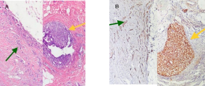
Background & objective: Breast cancer is thought to arise from non-invasive breast lesions, such as atypical ductal hyperplasia (ADH) and ductal carcinoma in situ (DCIS). DCIS is considered a direct precursor of invasive carcinoma. The morphological features alone do not reflect the biological truth of this disease. Therefore, we investigated features of carcinoma in situ and the invasive components in women diagnosed with breast cancer.
Methods: This study was a cross-sectional study. The corresponding IHC slides were selected from the pathology archive and examined by the pathologist. Fifty-one samples which showed both in situ and invasive components confirmed immunohistochemically, were included in the study.
Results: In 70.6% of the cases a high grade of in situ and invasive carcinoma was observed. In 45.1% of the studied cases, a solid structure was observed in in-situ carcinoma, and no otherwise specified structure was observed in invasive carcinoma. In 74.5% of both in situ and invasive carcinoma types, ER.PR had a positive value. In 45.5% of the cases, both in situ and invasive carcinoma components show low Ki67. In 42.2%, both in situ and invasive carcinomas were Her2 negative. There was no significant difference between the grade (P=0.687), Her2 type (P=0.532), and structure (P=0.532). ER.PR (P=1.00) and Ki67 (P=0.180) of in situ and invasive carcinoma in this study.
Conclusion: Our study showed differences between in situ and invasive biomarker expression. According to our findings, owing to heterogeneity, in situ components can't be representative of invasive components for treatment choices.

 求助内容:
求助内容: 应助结果提醒方式:


