Martina Manfredi, Simona Morabito, Quentin Fournier, Ioannis Panopoulos, Florence Thierry, Tobias Schwarz, Cristobal Lopez, Manuela Baldinetti, Chiara Massarenti, Davide Danilo Zani, Maurizio Longo
{"title":"卵巢肿瘤犬的ct表现:在复杂的腹部肿块中,弯曲的卵巢动脉一致地确定卵巢起源。","authors":"Martina Manfredi, Simona Morabito, Quentin Fournier, Ioannis Panopoulos, Florence Thierry, Tobias Schwarz, Cristobal Lopez, Manuela Baldinetti, Chiara Massarenti, Davide Danilo Zani, Maurizio Longo","doi":"10.1111/vru.13476","DOIUrl":null,"url":null,"abstract":"<p><p>The aim of this retrospective multicentric case series is to describe the CT findings of ovarian neoplasia in dogs. Twenty dogs with pre- and postcontrast CT exams and cytological/histological diagnosis of ovarian neoplasia were included. Five dogs presented with bilateral tumors, for a total of 25 neoplasms: 15 carcinomas (4 bilateral), 4 granulosa cell tumors, 2 poorly differentiated malignant neoplasia (bilateral), 2 luteomas, 1 teratoma, 1 dysgerminoma. In two dogs, the tumor developed from an ovarian remnant. Ovarian tumors showed variable size, lobulated shape, and precontrast heterogenous appearance. Mineral foci and/or fat components were rare, observed in teratoma, granulosa cell tumors (2), and ovarian carcinoma. Tumor type was not found to be associated with any CT features. Larger masses were more likely located in the central abdomen ventral to the ipsilateral kidney, demonstrated signs of tumor rupture, and were associated with abdominal or sternal lymphadenopathy and peritoneal effusion. A tortuous ovarian artery was constantly detectable, associated with an enlarged gonadal vein (12 cases). Related cavitary changes were peritoneal effusion (14 dogs) and sternal lymphadenopathy (7 dogs). Presumed or confirmed metastasis was reported in 9 of 20 cases, with CT evidence of transcoelomic (serosal thickening, peritoneal nodules, omental cake, implant lesions to the liver, spleen, and diaphragm), lymphatic and hematogenous spread (lungs, liver, bone, muscles, and spleen). In conclusion, the present study reports the CT features of different canine ovarian neoplasia. A tortuous ovarian artery may be useful to consistently recognize the ovarian origin of a large abdominal mass.</p>","PeriodicalId":23581,"journal":{"name":"Veterinary Radiology & Ultrasound","volume":"66 1","pages":"e13476"},"PeriodicalIF":1.5000,"publicationDate":"2025-01-01","publicationTypes":"Journal Article","fieldsOfStudy":null,"isOpenAccess":false,"openAccessPdf":"https://www.ncbi.nlm.nih.gov/pmc/articles/PMC11649852/pdf/","citationCount":"0","resultStr":"{\"title\":\"Computed tomographic findings in dogs with ovarian tumors: A tortuous ovarian artery consistently identifies ovarian origin in complex abdominal masses.\",\"authors\":\"Martina Manfredi, Simona Morabito, Quentin Fournier, Ioannis Panopoulos, Florence Thierry, Tobias Schwarz, Cristobal Lopez, Manuela Baldinetti, Chiara Massarenti, Davide Danilo Zani, Maurizio Longo\",\"doi\":\"10.1111/vru.13476\",\"DOIUrl\":null,\"url\":null,\"abstract\":\"<p><p>The aim of this retrospective multicentric case series is to describe the CT findings of ovarian neoplasia in dogs. Twenty dogs with pre- and postcontrast CT exams and cytological/histological diagnosis of ovarian neoplasia were included. Five dogs presented with bilateral tumors, for a total of 25 neoplasms: 15 carcinomas (4 bilateral), 4 granulosa cell tumors, 2 poorly differentiated malignant neoplasia (bilateral), 2 luteomas, 1 teratoma, 1 dysgerminoma. In two dogs, the tumor developed from an ovarian remnant. Ovarian tumors showed variable size, lobulated shape, and precontrast heterogenous appearance. Mineral foci and/or fat components were rare, observed in teratoma, granulosa cell tumors (2), and ovarian carcinoma. Tumor type was not found to be associated with any CT features. Larger masses were more likely located in the central abdomen ventral to the ipsilateral kidney, demonstrated signs of tumor rupture, and were associated with abdominal or sternal lymphadenopathy and peritoneal effusion. A tortuous ovarian artery was constantly detectable, associated with an enlarged gonadal vein (12 cases). Related cavitary changes were peritoneal effusion (14 dogs) and sternal lymphadenopathy (7 dogs). Presumed or confirmed metastasis was reported in 9 of 20 cases, with CT evidence of transcoelomic (serosal thickening, peritoneal nodules, omental cake, implant lesions to the liver, spleen, and diaphragm), lymphatic and hematogenous spread (lungs, liver, bone, muscles, and spleen). In conclusion, the present study reports the CT features of different canine ovarian neoplasia. A tortuous ovarian artery may be useful to consistently recognize the ovarian origin of a large abdominal mass.</p>\",\"PeriodicalId\":23581,\"journal\":{\"name\":\"Veterinary Radiology & Ultrasound\",\"volume\":\"66 1\",\"pages\":\"e13476\"},\"PeriodicalIF\":1.5000,\"publicationDate\":\"2025-01-01\",\"publicationTypes\":\"Journal Article\",\"fieldsOfStudy\":null,\"isOpenAccess\":false,\"openAccessPdf\":\"https://www.ncbi.nlm.nih.gov/pmc/articles/PMC11649852/pdf/\",\"citationCount\":\"0\",\"resultStr\":null,\"platform\":\"Semanticscholar\",\"paperid\":null,\"PeriodicalName\":\"Veterinary Radiology & Ultrasound\",\"FirstCategoryId\":\"97\",\"ListUrlMain\":\"https://doi.org/10.1111/vru.13476\",\"RegionNum\":2,\"RegionCategory\":\"农林科学\",\"ArticlePicture\":[],\"TitleCN\":null,\"AbstractTextCN\":null,\"PMCID\":null,\"EPubDate\":\"\",\"PubModel\":\"\",\"JCR\":\"Q2\",\"JCRName\":\"VETERINARY SCIENCES\",\"Score\":null,\"Total\":0}","platform":"Semanticscholar","paperid":null,"PeriodicalName":"Veterinary Radiology & Ultrasound","FirstCategoryId":"97","ListUrlMain":"https://doi.org/10.1111/vru.13476","RegionNum":2,"RegionCategory":"农林科学","ArticlePicture":[],"TitleCN":null,"AbstractTextCN":null,"PMCID":null,"EPubDate":"","PubModel":"","JCR":"Q2","JCRName":"VETERINARY SCIENCES","Score":null,"Total":0}
Computed tomographic findings in dogs with ovarian tumors: A tortuous ovarian artery consistently identifies ovarian origin in complex abdominal masses.
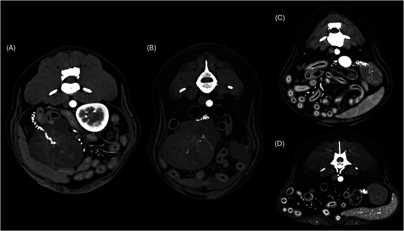
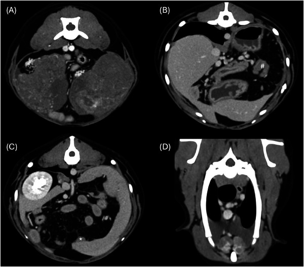
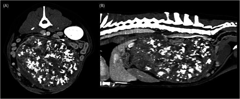
The aim of this retrospective multicentric case series is to describe the CT findings of ovarian neoplasia in dogs. Twenty dogs with pre- and postcontrast CT exams and cytological/histological diagnosis of ovarian neoplasia were included. Five dogs presented with bilateral tumors, for a total of 25 neoplasms: 15 carcinomas (4 bilateral), 4 granulosa cell tumors, 2 poorly differentiated malignant neoplasia (bilateral), 2 luteomas, 1 teratoma, 1 dysgerminoma. In two dogs, the tumor developed from an ovarian remnant. Ovarian tumors showed variable size, lobulated shape, and precontrast heterogenous appearance. Mineral foci and/or fat components were rare, observed in teratoma, granulosa cell tumors (2), and ovarian carcinoma. Tumor type was not found to be associated with any CT features. Larger masses were more likely located in the central abdomen ventral to the ipsilateral kidney, demonstrated signs of tumor rupture, and were associated with abdominal or sternal lymphadenopathy and peritoneal effusion. A tortuous ovarian artery was constantly detectable, associated with an enlarged gonadal vein (12 cases). Related cavitary changes were peritoneal effusion (14 dogs) and sternal lymphadenopathy (7 dogs). Presumed or confirmed metastasis was reported in 9 of 20 cases, with CT evidence of transcoelomic (serosal thickening, peritoneal nodules, omental cake, implant lesions to the liver, spleen, and diaphragm), lymphatic and hematogenous spread (lungs, liver, bone, muscles, and spleen). In conclusion, the present study reports the CT features of different canine ovarian neoplasia. A tortuous ovarian artery may be useful to consistently recognize the ovarian origin of a large abdominal mass.
期刊介绍:
Veterinary Radiology & Ultrasound is a bimonthly, international, peer-reviewed, research journal devoted to the fields of veterinary diagnostic imaging and radiation oncology. Established in 1958, it is owned by the American College of Veterinary Radiology and is also the official journal for six affiliate veterinary organizations. Veterinary Radiology & Ultrasound is represented on the International Committee of Medical Journal Editors, World Association of Medical Editors, and Committee on Publication Ethics.
The mission of Veterinary Radiology & Ultrasound is to serve as a leading resource for high quality articles that advance scientific knowledge and standards of clinical practice in the areas of veterinary diagnostic radiology, computed tomography, magnetic resonance imaging, ultrasonography, nuclear imaging, radiation oncology, and interventional radiology. Manuscript types include original investigations, imaging diagnosis reports, review articles, editorials and letters to the Editor. Acceptance criteria include originality, significance, quality, reader interest, composition and adherence to author guidelines.

 求助内容:
求助内容: 应助结果提醒方式:


