{"title":"自发性寄生带蒂肌瘤,表现为子宫平滑肌细胞缺失1例。","authors":"Naoki Shibata, Michihisa Shiro, Noriyoshi Oki, Takahiro Watanabe, Hitomi Futaki, Shigeki Yoshida","doi":"10.4103/gmit.gmit_151_23","DOIUrl":null,"url":null,"abstract":"<p><p>A few cases of spontaneous parasitic myoma have been reported. However, its cause remains unidentified. We report a case of spontaneous parasitic pedunculated subserosal myoma with pathological findings presenting with the absence of uterine smooth muscle cells in the stalk observed during robotic-assisted laparoscopic hysterectomy. A 51-year-old patient (G1P0) with no prior surgical history underwent a robotic-assisted laparoscopic hysterectomy. An approximately 3 cm-pedunculated subserosal myoma was found attached to the retroperitoneum. The stalk was sealed and separated and the myoma with retroperitoneal adipose tissue was resected. The stalk was pathologically identified to lack uterine smooth muscle cells and contain only muscular arteries and fibrous connective tissues. Thus, it might be hypothesized that after the myoma received collateral parasitic blood flow from the attached retroperitoneum, the stalk degenerated, and uterine smooth muscle cells were lost through an unknown mechanism, possibly underlying the development of spontaneous parasitic myomas.</p>","PeriodicalId":45272,"journal":{"name":"Gynecology and Minimally Invasive Therapy-GMIT","volume":"13 4","pages":"265-268"},"PeriodicalIF":1.7000,"publicationDate":"2024-10-21","publicationTypes":"Journal Article","fieldsOfStudy":null,"isOpenAccess":false,"openAccessPdf":"https://www.ncbi.nlm.nih.gov/pmc/articles/PMC11626902/pdf/","citationCount":"0","resultStr":"{\"title\":\"Spontaneous Parasitic Pedunculated Myoma Presenting the Absence of Uterine Smooth Muscle Cells in the Stalk - A Case Report.\",\"authors\":\"Naoki Shibata, Michihisa Shiro, Noriyoshi Oki, Takahiro Watanabe, Hitomi Futaki, Shigeki Yoshida\",\"doi\":\"10.4103/gmit.gmit_151_23\",\"DOIUrl\":null,\"url\":null,\"abstract\":\"<p><p>A few cases of spontaneous parasitic myoma have been reported. However, its cause remains unidentified. We report a case of spontaneous parasitic pedunculated subserosal myoma with pathological findings presenting with the absence of uterine smooth muscle cells in the stalk observed during robotic-assisted laparoscopic hysterectomy. A 51-year-old patient (G1P0) with no prior surgical history underwent a robotic-assisted laparoscopic hysterectomy. An approximately 3 cm-pedunculated subserosal myoma was found attached to the retroperitoneum. The stalk was sealed and separated and the myoma with retroperitoneal adipose tissue was resected. The stalk was pathologically identified to lack uterine smooth muscle cells and contain only muscular arteries and fibrous connective tissues. Thus, it might be hypothesized that after the myoma received collateral parasitic blood flow from the attached retroperitoneum, the stalk degenerated, and uterine smooth muscle cells were lost through an unknown mechanism, possibly underlying the development of spontaneous parasitic myomas.</p>\",\"PeriodicalId\":45272,\"journal\":{\"name\":\"Gynecology and Minimally Invasive Therapy-GMIT\",\"volume\":\"13 4\",\"pages\":\"265-268\"},\"PeriodicalIF\":1.7000,\"publicationDate\":\"2024-10-21\",\"publicationTypes\":\"Journal Article\",\"fieldsOfStudy\":null,\"isOpenAccess\":false,\"openAccessPdf\":\"https://www.ncbi.nlm.nih.gov/pmc/articles/PMC11626902/pdf/\",\"citationCount\":\"0\",\"resultStr\":null,\"platform\":\"Semanticscholar\",\"paperid\":null,\"PeriodicalName\":\"Gynecology and Minimally Invasive Therapy-GMIT\",\"FirstCategoryId\":\"1085\",\"ListUrlMain\":\"https://doi.org/10.4103/gmit.gmit_151_23\",\"RegionNum\":0,\"RegionCategory\":null,\"ArticlePicture\":[],\"TitleCN\":null,\"AbstractTextCN\":null,\"PMCID\":null,\"EPubDate\":\"2024/10/1 0:00:00\",\"PubModel\":\"eCollection\",\"JCR\":\"Q3\",\"JCRName\":\"OBSTETRICS & GYNECOLOGY\",\"Score\":null,\"Total\":0}","platform":"Semanticscholar","paperid":null,"PeriodicalName":"Gynecology and Minimally Invasive Therapy-GMIT","FirstCategoryId":"1085","ListUrlMain":"https://doi.org/10.4103/gmit.gmit_151_23","RegionNum":0,"RegionCategory":null,"ArticlePicture":[],"TitleCN":null,"AbstractTextCN":null,"PMCID":null,"EPubDate":"2024/10/1 0:00:00","PubModel":"eCollection","JCR":"Q3","JCRName":"OBSTETRICS & GYNECOLOGY","Score":null,"Total":0}
Spontaneous Parasitic Pedunculated Myoma Presenting the Absence of Uterine Smooth Muscle Cells in the Stalk - A Case Report.
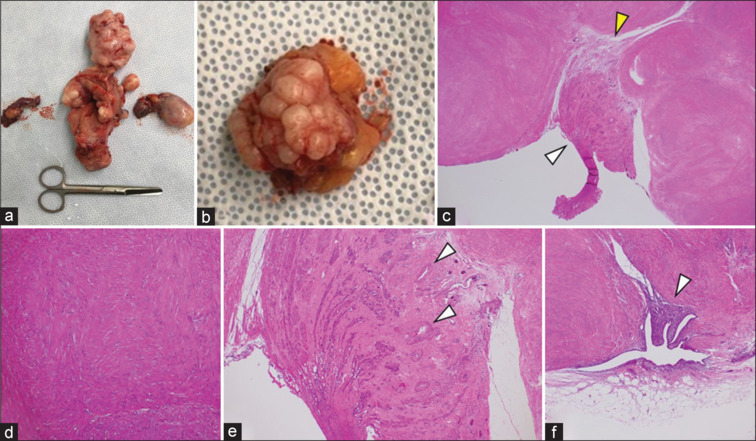
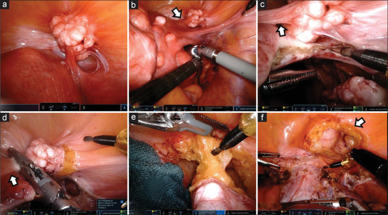
A few cases of spontaneous parasitic myoma have been reported. However, its cause remains unidentified. We report a case of spontaneous parasitic pedunculated subserosal myoma with pathological findings presenting with the absence of uterine smooth muscle cells in the stalk observed during robotic-assisted laparoscopic hysterectomy. A 51-year-old patient (G1P0) with no prior surgical history underwent a robotic-assisted laparoscopic hysterectomy. An approximately 3 cm-pedunculated subserosal myoma was found attached to the retroperitoneum. The stalk was sealed and separated and the myoma with retroperitoneal adipose tissue was resected. The stalk was pathologically identified to lack uterine smooth muscle cells and contain only muscular arteries and fibrous connective tissues. Thus, it might be hypothesized that after the myoma received collateral parasitic blood flow from the attached retroperitoneum, the stalk degenerated, and uterine smooth muscle cells were lost through an unknown mechanism, possibly underlying the development of spontaneous parasitic myomas.

 求助内容:
求助内容: 应助结果提醒方式:


