Ilenia Di Cola, Léa Savey, Marion Delplanque, Rim Bourguiba, Alessandra Bartoli, Zohra Aknouche, Fatima Bensalek, Isabelle Kone‐Paut, Linda Rossi‐Semerano, Isabelle Melki, Brigitte Bader‐Meunier, Piero Ruscitti, Bénédicte Neven, Pierre Quartier, Guilaine Boursier, Irina Giurgea, Laurence Cuisset, Gilles Grateau, Véronique Hentgen, Sophie Georgin‐Lavialle
{"title":"家族性地中海热缺铁:来自JIR队列的211名成年患者的研究","authors":"Ilenia Di Cola, Léa Savey, Marion Delplanque, Rim Bourguiba, Alessandra Bartoli, Zohra Aknouche, Fatima Bensalek, Isabelle Kone‐Paut, Linda Rossi‐Semerano, Isabelle Melki, Brigitte Bader‐Meunier, Piero Ruscitti, Bénédicte Neven, Pierre Quartier, Guilaine Boursier, Irina Giurgea, Laurence Cuisset, Gilles Grateau, Véronique Hentgen, Sophie Georgin‐Lavialle","doi":"10.1002/ajh.27549","DOIUrl":null,"url":null,"abstract":"<p>Familial Mediterranean fever (FMF) is the most common monogenic autoinflammatory disease worldwide associated with <i>MEFV</i> mutations. Patients display recurrent and self-limited attacks of fever, abdominal and thoracic pain [<span>1</span>]. There is no specific association between anemia and FMF, except that patients with chronic inflammation may have inflammatory microcytic anemia [<span>2</span>]. However, chronic anemia can lead to fatigue, and fatigue is known to be a trigger for FMF attacks [<span>2, 5</span>]. Therefore, patients with fatigue due to anemia may have more flare-ups of the disease and worse quality of life [<span>2</span>]. Iron deficiency can cause fatigue even in the absence of anemia [<span>3</span>]. Fatigue is also commonly reported in FMF [<span>2</span>]. Therefore, it may be beneficial to look for iron deficiency without anemia as one of the curable causes of fatigue in FMF, especially as fatigue may be considered a trigger of their attacks.</p>\n<p>Although a variety of laboratory assessments of iron status is available, serum ferritin, a circulating protein in equilibrium with tissue ferritin, is the most sensitive and specific test for detecting isolated iron deficiency [<span>4</span>]. However, the assessment of serum ferritin could be biased by the inflammatory background in an acute inflammatory attack of such an autoinflammatory disease because in case of a normal or high rate it may not inform on the real absence of iron deficiency.</p>\n<p>Our aim was to assess the prevalence of iron deficiency in FMF and its association with clinical features, laboratory parameters, and therapies.</p>\n<p>From 2016 to 2023, a retrospective evaluation of prospectively followed FMF patients at the French national FMF-reference center was performed to analyze the prevalence of iron deficiency and its association with clinical features, laboratory parameters, and therapies. The presence of iron deficiency was defined according to the ferritin threshold of our hospital laboratory (ferritin < 27.0 ng/mL), thus, homogeneously collected and measured. Ferritin levels were collected from medical records of follow-up visits. Considering the retrospective nature of our study, we collected what reported in clinical chart of patients; however, some relevant parameters (i.e., transferrin saturation, serum transferrin receptor levels) were not included in the analyses since they are not performed in routine care.</p>\n<p>All FMF displayed two pathogenic exon 10 <i>MEFV</i> mutations either M694V (pMet694Val) and/or M694I (pMet694Ile). After collection of informed consents, we extracted data from the Juvenile Inflammatory Rheumatism (JIR) cohort, an international multicentric data repository authorized by the National Commission on Informatics and Liberties (CNIL, authorization number 914677).</p>\n<p>For the statistics, clinical features were exploratively compared, according to the presence of iron deficiency (ferritin < 27.0 ng/mL) by <i>t</i>-tests for continuous or categorical variables, as appropriate. After, multivariate regression models were built to exploit the clinical risk profile of patients with ferritin < 27.0 ng/mL. The selection process of variables started by a univariate analysis; any variable having a significant univariate test was tested as a possible candidate for the multivariate analysis. Additionally, variables were tested for multivariate analysis according to their clinical relevance. At the end of this multistep process, we built multivariate models, providing OR estimations of the clinical risk profile of patients with ferritin < 27.0 ng/mL. A <i>p</i> value < 0.05 was considered statistically significant. Patients with missing data were excluded from the analysis. The Statistics Package for Social Sciences (SPSS version 17.0, SPSS Inc.) was used for all analyses.</p>\n<p>Overall, 211 FMF were included, mostly female (60.2%) and with a mean age of 41.34 [18–89] years. Out of 208 patients with the value of ferritin reported in our database, 67 (31.80%) had a serum ferritin level < 27 ng/mL and were defined as having iron deficiency. Among these, 61 (91.04%) were female and with a mean age of 36.81 [18–89] years. Among the 67 with iron deficiency, 42 (62.69%) displayed anemia (Hb < 13.0 g/dL), including 40 female and 2 males. Patients with iron deficiency showed lower values of Hb (<i>p</i> < 0.001), mean corpuscular volume (MCV) (<i>p</i> = 0.002), BMI (<i>p</i> = 0.044), weight (<i>p</i> < 0.001), daily colchicine dose (<i>p</i> = 0.015), and creatinine (<i>p</i> = 0.005) and they were younger than those without iron deficiency (<i>p</i> = 0.011). No differences were retrieved comparing these two groups regarding inflammatory markers. Particularly, 52.24% of FMF with ferritin < 27 showed normal inflammatory markers (Table S1). Among the 82 men, 6 showed a serum ferritin level < 27 ng/mL. These patients did not significantly differ in main features compared with those without iron deficiency (Table S2).</p>\n<p>The difference between values of Hb and daily colchicine dose according to iron deficiency were also analyzed (Figure S1).</p>\n<p>In our cohort, both univariate and multivariate regression models were built to evaluate the clinical risk profile of patients with ferritin < 27 ng/mL (Table 1). Based on univariate analyses, clinical relevance but also considering the number of patients characterized by ferritin < 27 ng/mL, we built two multivariate regression models. One model comprised age, female sex, CRP, SAA, BMI, and colchicine doses whereas the other included age, female sex, CRP, SAA, colchicine doses, and biological disease-modifying antirheumatic drugs (bDMARDs). Despite not significant univariate analysis, CRP and SAA were added in the models considering the possible influence of the inflammatory process on ferritin levels [<span>6</span>]. We added bDMARDs in a model considering that more severe patients were probably treated with these drugs. In the first model, age (OR: 0.97, 95% CI: 0.95–1.00, <i>p</i> = 0.041) and female sex (OR: 13.77, 95% CI: 4.96–38.18, <i>p</i> < 0.001) were significantly and independently associated with the presence of ferritin < 27 ng/mL. Similarly, in the other model, age (OR: 0.97, 95% CI: 0.95–1.00, <i>p</i> = 0.015), and female sex (OR: 16.15, 95% CI: 5.67–46.00, <i>p</i> < 0.001) significantly predicted ferritin < 27 ng/mL.</p>\n<div>\n<header><span>TABLE 1. </span>Regression analyses evaluating the clinical risk profile of patients characterized by ferritin < 27 ng/mL.</header>\n<div tabindex=\"0\">\n<table>\n<thead>\n<tr>\n<th>Clinical variables</th>\n<th>OR</th>\n<th>SE</th>\n<th>95% CI</th>\n<th>\n<i>p</i>\n</th>\n</tr>\n</thead>\n<tbody>\n<tr>\n<td colspan=\"5\">\n<b>Ferritin < 27 ng/mL</b>\n</td>\n</tr>\n<tr>\n<td colspan=\"5\">Univariate analysis</td>\n</tr>\n<tr>\n<td style=\"padding-left:2em;\">Age</td>\n<td>0.98</td>\n<td>0.009</td>\n<td>0.95–1.00</td>\n<td>0.063</td>\n</tr>\n<tr>\n<td style=\"padding-left:2em;\">Female sex</td>\n<td>11.89</td>\n<td>0.460</td>\n<td>4.83–29.28</td>\n<td>\n<b>< 0.001</b>\n</td>\n</tr>\n<tr>\n<td style=\"padding-left:2em;\">Hb</td>\n<td>0.62</td>\n<td>0.099</td>\n<td>0.51–0.75</td>\n<td>\n<b>< 0.001</b>\n</td>\n</tr>\n<tr>\n<td style=\"padding-left:2em;\">Hb < 13.0 g/dL</td>\n<td>3.01</td>\n<td>0.310</td>\n<td>1.64–5.53</td>\n<td>\n<b>< 0.001</b>\n</td>\n</tr>\n<tr>\n<td style=\"padding-left:2em;\">RBC</td>\n<td>0.47</td>\n<td>0.271</td>\n<td>0.28–0.81</td>\n<td>\n<b>0.006</b>\n</td>\n</tr>\n<tr>\n<td style=\"padding-left:2em;\">MCV</td>\n<td>0.89</td>\n<td>0.036</td>\n<td>0.83–0.96</td>\n<td>\n<b>0.002</b>\n</td>\n</tr>\n<tr>\n<td style=\"padding-left:2em;\">CRP</td>\n<td>0.98</td>\n<td>0.010</td>\n<td>0.96–1.00</td>\n<td>0.088</td>\n</tr>\n<tr>\n<td style=\"padding-left:2em;\">SAA</td>\n<td>0.99</td>\n<td>0.003</td>\n<td>0.99–1.00</td>\n<td>0.288</td>\n</tr>\n<tr>\n<td style=\"padding-left:2em;\">BMI</td>\n<td>0.93</td>\n<td>0.037</td>\n<td>0.87–0.99</td>\n<td>\n<b>0.045</b>\n</td>\n</tr>\n<tr>\n<td style=\"padding-left:2em;\">Daily colchicine dose</td>\n<td>1.87</td>\n<td>0.261</td>\n<td>1.12–3.11</td>\n<td>\n<b>0.016</b>\n</td>\n</tr>\n<tr>\n<td style=\"padding-left:2em;\">bDMARDs</td>\n<td>0.74</td>\n<td>0.377</td>\n<td>0.36–1.56</td>\n<td>0.431</td>\n</tr>\n<tr>\n<td style=\"padding-left:2em;\">Weight</td>\n<td>0.96</td>\n<td>0.013</td>\n<td>0.93–0.98</td>\n<td>\n<b>< 0.001</b>\n</td>\n</tr>\n<tr>\n<td style=\"padding-left:2em;\">Creatinine</td>\n<td>0.94</td>\n<td>0.014</td>\n<td>0.91–0.97</td>\n<td>\n<b>< 0.001</b>\n</td>\n</tr>\n<tr>\n<td colspan=\"5\">Multivariate analysis</td>\n</tr>\n<tr>\n<td style=\"padding-left:2em;\">Age</td>\n<td>0.97</td>\n<td>0.013</td>\n<td>0.95–1.00</td>\n<td>\n<b>0.041</b>\n</td>\n</tr>\n<tr>\n<td style=\"padding-left:2em;\">Female sex</td>\n<td>13.77</td>\n<td>0.521</td>\n<td>4.96–38.18</td>\n<td>\n<b>< 0.001</b>\n</td>\n</tr>\n<tr>\n<td style=\"padding-left:2em;\">CRP</td>\n<td>0.99</td>\n<td>0.014</td>\n<td>0.96–1.01</td>\n<td>0.325</td>\n</tr>\n<tr>\n<td style=\"padding-left:2em;\">SAA</td>\n<td>1.00</td>\n<td>0.003</td>\n<td>0.99–1.00</td>\n<td>0.680</td>\n</tr>\n<tr>\n<td style=\"padding-left:2em;\">BMI</td>\n<td>0.990</td>\n<td>0.044</td>\n<td>0.91–1.08</td>\n<td>0.815</td>\n</tr>\n<tr>\n<td style=\"padding-left:2em;\">bDMARDs</td>\n<td>1.56</td>\n<td>0.325</td>\n<td>0.83–2.96</td>\n<td>0.170</td>\n</tr>\n<tr>\n<td colspan=\"5\">Multivariate analysis</td>\n</tr>\n<tr>\n<td style=\"padding-left:2em;\">Age</td>\n<td>0.97</td>\n<td>0.013</td>\n<td>0.95–1.00</td>\n<td>\n<b>0.015</b>\n</td>\n</tr>\n<tr>\n<td style=\"padding-left:2em;\">Female sex</td>\n<td>16.15</td>\n<td>0.53</td>\n<td>5.67–46.00</td>\n<td>\n<b>< 0.001</b>\n</td>\n</tr>\n<tr>\n<td style=\"padding-left:2em;\">CRP</td>\n<td>0.99</td>\n<td>0.013</td>\n<td>0.96–1.01</td>\n<td>0.320</td>\n</tr>\n<tr>\n<td style=\"padding-left:2em;\">SAA</td>\n<td>1.00</td>\n<td>0.003</td>\n<td>0.99–1.00</td>\n<td>0.658</td>\n</tr>\n<tr>\n<td style=\"padding-left:2em;\">Daily colchicine dose</td>\n<td>1.80</td>\n<td>0.33</td>\n<td>0.94–3.47</td>\n<td>0.078</td>\n</tr>\n<tr>\n<td style=\"padding-left:2em;\">bDMARDs</td>\n<td>1.67</td>\n<td>0.52</td>\n<td>0.61–4.63</td>\n<td>\n<b>0.322</b>\n</td>\n</tr>\n</tbody>\n</table>\n</div>\n<div>\n<ul>\n<li>\n<i>Note: p</i> < 0.05 was considered statistically significant. </li>\n<li> Abbreviations: bDMARDs: biological disease-modifying antirheumatic drugs; BMI: body mass index; CRP: C reactive protein; Hb: hemoglobin; M: men; MCV: mean corpuscular volume; <i>N</i>: number of patients; SAA: serum amyloid A; SD: standard deviation; W: women. </li>\n</ul>\n</div>\n<div></div>\n</div>\n<p>Altogether, 31.8% of FMF displayed ferritin < 27 ng/mL and were mostly female and of younger age. Ferritin may be considered the mainstay for iron deficiency detection [<span>3</span>], which has been shown to be responsible for fatigue, a very frequent favoring factor in FMF flare-ups [<span>2</span>]. Although in absence of anemia, iron deficiency could be an additional factor in the onset of inflammatory flare-ups and may affect the patient's quality of life, although this aspect could not be fully studied retrospectively. Indeed, to clearly define an “iron deficiency” condition in FMF, also in absence of anemia, future studies are needed to assess other parameters of iron metabolism besides ferritin.</p>\n<p>Based on clinical observations, and the predominance of young female, regardless of their inflammatory status, our first hypothesis is that ferritin < 27 ng/mL and associated iron deficiency, also in absence of anemia, may be secondary to excessive gynecological loss not entirely compensated by an increased intake of iron-rich foods. Due to the retrospective nature of this study and the fact that information on the characteristics of the patients' periods was not systematically collected, an exhaustive answer to this question could not be given, but when the affected women were interviewed, they most often reported heavy and long-lasting periods without taking contraceptive pill. It is also possible that young women eat less animal protein. Finally, digestive bleeding, potentially aggravated by frequent use of NSAIDs, should not be overlooked. Iron deficiency did not cause anemia in all patients, but it can cause fatigue, and eventually FMF attacks, suggesting the needing of iron supplementation.</p>\n<p>Here, we showed that FMF patients with ferritin < 27 ng/mL had lower Hb and BMI and were significantly younger than others, without elevated inflammatory biomarkers. Interestingly, they required higher doses of colchicine, mainly 2 mg/day. The role of colchicine in decreasing iron intestinal absorption may be questioned. Our work does not allow us to answer this question, but not all patients with iron deficiency received the maximum daily colchicine dose.</p>\n<p>In our opinion, it is important to correct iron deficiency in FMF, starting with treating the cause of the iron loss and correcting the oral intake if possible and tolerated. Oral iron combined with oral colchicine may cause digestive discomfort and transit disorders, leading patients to stop iron supplements. If oral supplementation is not possible, intravenous iron infusions can be offered. In addition to iron supplements, patients can be given a list of iron-rich foods. Finding the cause of the martial deficiency with a particular focus on excessive gynecological bleeding to reduce the frequency and duration of menstruation in women should be done. It is also important to look for associated gastric disorders, especially in this population with a probable excess risk, or <i>Helicobacter pylori</i> infection. Our study should be considered as “hypothesis generating” to provide the basis for further specific designed studies to better elucidate this issue. To the best of our knowledge, this would be the first analysis of value of ferritin in FMF. This could be of importance to evaluate the complicated mechanisms underlying the asthenia as trigger of an inflammatory disease. We underline that use the ferritin alone as the sole arbiter of iron deficiency has to be confirmed especially among patients with a monogenic autoinflammatory diseases such as FMF if they display inflammation.</p>\n<p>This work highlights the importance of measuring ferritin levels in FMF, especially because this dosage is simple and available worldwide in routine care including some emerging countries where FMF is highly prevalent. In the presence of significant iron deficiency, iron supplementation should be done to correct it and reduce any iron deficiency signs such as asthenia and anemia that may lead to FMF attacks and also entertain an asthenia circle in FMF patients.</p>","PeriodicalId":7724,"journal":{"name":"American Journal of Hematology","volume":"35 1","pages":""},"PeriodicalIF":10.1000,"publicationDate":"2024-12-07","publicationTypes":"Journal Article","fieldsOfStudy":null,"isOpenAccess":false,"openAccessPdf":"","citationCount":"0","resultStr":"{\"title\":\"Iron Deficiency in Familial Mediterranean Fever: A Study on 211 Adult Patients From the JIR Cohort\",\"authors\":\"Ilenia Di Cola, Léa Savey, Marion Delplanque, Rim Bourguiba, Alessandra Bartoli, Zohra Aknouche, Fatima Bensalek, Isabelle Kone‐Paut, Linda Rossi‐Semerano, Isabelle Melki, Brigitte Bader‐Meunier, Piero Ruscitti, Bénédicte Neven, Pierre Quartier, Guilaine Boursier, Irina Giurgea, Laurence Cuisset, Gilles Grateau, Véronique Hentgen, Sophie Georgin‐Lavialle\",\"doi\":\"10.1002/ajh.27549\",\"DOIUrl\":null,\"url\":null,\"abstract\":\"<p>Familial Mediterranean fever (FMF) is the most common monogenic autoinflammatory disease worldwide associated with <i>MEFV</i> mutations. Patients display recurrent and self-limited attacks of fever, abdominal and thoracic pain [<span>1</span>]. There is no specific association between anemia and FMF, except that patients with chronic inflammation may have inflammatory microcytic anemia [<span>2</span>]. However, chronic anemia can lead to fatigue, and fatigue is known to be a trigger for FMF attacks [<span>2, 5</span>]. Therefore, patients with fatigue due to anemia may have more flare-ups of the disease and worse quality of life [<span>2</span>]. Iron deficiency can cause fatigue even in the absence of anemia [<span>3</span>]. Fatigue is also commonly reported in FMF [<span>2</span>]. Therefore, it may be beneficial to look for iron deficiency without anemia as one of the curable causes of fatigue in FMF, especially as fatigue may be considered a trigger of their attacks.</p>\\n<p>Although a variety of laboratory assessments of iron status is available, serum ferritin, a circulating protein in equilibrium with tissue ferritin, is the most sensitive and specific test for detecting isolated iron deficiency [<span>4</span>]. However, the assessment of serum ferritin could be biased by the inflammatory background in an acute inflammatory attack of such an autoinflammatory disease because in case of a normal or high rate it may not inform on the real absence of iron deficiency.</p>\\n<p>Our aim was to assess the prevalence of iron deficiency in FMF and its association with clinical features, laboratory parameters, and therapies.</p>\\n<p>From 2016 to 2023, a retrospective evaluation of prospectively followed FMF patients at the French national FMF-reference center was performed to analyze the prevalence of iron deficiency and its association with clinical features, laboratory parameters, and therapies. The presence of iron deficiency was defined according to the ferritin threshold of our hospital laboratory (ferritin < 27.0 ng/mL), thus, homogeneously collected and measured. Ferritin levels were collected from medical records of follow-up visits. Considering the retrospective nature of our study, we collected what reported in clinical chart of patients; however, some relevant parameters (i.e., transferrin saturation, serum transferrin receptor levels) were not included in the analyses since they are not performed in routine care.</p>\\n<p>All FMF displayed two pathogenic exon 10 <i>MEFV</i> mutations either M694V (pMet694Val) and/or M694I (pMet694Ile). After collection of informed consents, we extracted data from the Juvenile Inflammatory Rheumatism (JIR) cohort, an international multicentric data repository authorized by the National Commission on Informatics and Liberties (CNIL, authorization number 914677).</p>\\n<p>For the statistics, clinical features were exploratively compared, according to the presence of iron deficiency (ferritin < 27.0 ng/mL) by <i>t</i>-tests for continuous or categorical variables, as appropriate. After, multivariate regression models were built to exploit the clinical risk profile of patients with ferritin < 27.0 ng/mL. The selection process of variables started by a univariate analysis; any variable having a significant univariate test was tested as a possible candidate for the multivariate analysis. Additionally, variables were tested for multivariate analysis according to their clinical relevance. At the end of this multistep process, we built multivariate models, providing OR estimations of the clinical risk profile of patients with ferritin < 27.0 ng/mL. A <i>p</i> value < 0.05 was considered statistically significant. Patients with missing data were excluded from the analysis. The Statistics Package for Social Sciences (SPSS version 17.0, SPSS Inc.) was used for all analyses.</p>\\n<p>Overall, 211 FMF were included, mostly female (60.2%) and with a mean age of 41.34 [18–89] years. Out of 208 patients with the value of ferritin reported in our database, 67 (31.80%) had a serum ferritin level < 27 ng/mL and were defined as having iron deficiency. Among these, 61 (91.04%) were female and with a mean age of 36.81 [18–89] years. Among the 67 with iron deficiency, 42 (62.69%) displayed anemia (Hb < 13.0 g/dL), including 40 female and 2 males. Patients with iron deficiency showed lower values of Hb (<i>p</i> < 0.001), mean corpuscular volume (MCV) (<i>p</i> = 0.002), BMI (<i>p</i> = 0.044), weight (<i>p</i> < 0.001), daily colchicine dose (<i>p</i> = 0.015), and creatinine (<i>p</i> = 0.005) and they were younger than those without iron deficiency (<i>p</i> = 0.011). No differences were retrieved comparing these two groups regarding inflammatory markers. Particularly, 52.24% of FMF with ferritin < 27 showed normal inflammatory markers (Table S1). Among the 82 men, 6 showed a serum ferritin level < 27 ng/mL. These patients did not significantly differ in main features compared with those without iron deficiency (Table S2).</p>\\n<p>The difference between values of Hb and daily colchicine dose according to iron deficiency were also analyzed (Figure S1).</p>\\n<p>In our cohort, both univariate and multivariate regression models were built to evaluate the clinical risk profile of patients with ferritin < 27 ng/mL (Table 1). Based on univariate analyses, clinical relevance but also considering the number of patients characterized by ferritin < 27 ng/mL, we built two multivariate regression models. One model comprised age, female sex, CRP, SAA, BMI, and colchicine doses whereas the other included age, female sex, CRP, SAA, colchicine doses, and biological disease-modifying antirheumatic drugs (bDMARDs). Despite not significant univariate analysis, CRP and SAA were added in the models considering the possible influence of the inflammatory process on ferritin levels [<span>6</span>]. We added bDMARDs in a model considering that more severe patients were probably treated with these drugs. In the first model, age (OR: 0.97, 95% CI: 0.95–1.00, <i>p</i> = 0.041) and female sex (OR: 13.77, 95% CI: 4.96–38.18, <i>p</i> < 0.001) were significantly and independently associated with the presence of ferritin < 27 ng/mL. Similarly, in the other model, age (OR: 0.97, 95% CI: 0.95–1.00, <i>p</i> = 0.015), and female sex (OR: 16.15, 95% CI: 5.67–46.00, <i>p</i> < 0.001) significantly predicted ferritin < 27 ng/mL.</p>\\n<div>\\n<header><span>TABLE 1. </span>Regression analyses evaluating the clinical risk profile of patients characterized by ferritin < 27 ng/mL.</header>\\n<div tabindex=\\\"0\\\">\\n<table>\\n<thead>\\n<tr>\\n<th>Clinical variables</th>\\n<th>OR</th>\\n<th>SE</th>\\n<th>95% CI</th>\\n<th>\\n<i>p</i>\\n</th>\\n</tr>\\n</thead>\\n<tbody>\\n<tr>\\n<td colspan=\\\"5\\\">\\n<b>Ferritin < 27 ng/mL</b>\\n</td>\\n</tr>\\n<tr>\\n<td colspan=\\\"5\\\">Univariate analysis</td>\\n</tr>\\n<tr>\\n<td style=\\\"padding-left:2em;\\\">Age</td>\\n<td>0.98</td>\\n<td>0.009</td>\\n<td>0.95–1.00</td>\\n<td>0.063</td>\\n</tr>\\n<tr>\\n<td style=\\\"padding-left:2em;\\\">Female sex</td>\\n<td>11.89</td>\\n<td>0.460</td>\\n<td>4.83–29.28</td>\\n<td>\\n<b>< 0.001</b>\\n</td>\\n</tr>\\n<tr>\\n<td style=\\\"padding-left:2em;\\\">Hb</td>\\n<td>0.62</td>\\n<td>0.099</td>\\n<td>0.51–0.75</td>\\n<td>\\n<b>< 0.001</b>\\n</td>\\n</tr>\\n<tr>\\n<td style=\\\"padding-left:2em;\\\">Hb < 13.0 g/dL</td>\\n<td>3.01</td>\\n<td>0.310</td>\\n<td>1.64–5.53</td>\\n<td>\\n<b>< 0.001</b>\\n</td>\\n</tr>\\n<tr>\\n<td style=\\\"padding-left:2em;\\\">RBC</td>\\n<td>0.47</td>\\n<td>0.271</td>\\n<td>0.28–0.81</td>\\n<td>\\n<b>0.006</b>\\n</td>\\n</tr>\\n<tr>\\n<td style=\\\"padding-left:2em;\\\">MCV</td>\\n<td>0.89</td>\\n<td>0.036</td>\\n<td>0.83–0.96</td>\\n<td>\\n<b>0.002</b>\\n</td>\\n</tr>\\n<tr>\\n<td style=\\\"padding-left:2em;\\\">CRP</td>\\n<td>0.98</td>\\n<td>0.010</td>\\n<td>0.96–1.00</td>\\n<td>0.088</td>\\n</tr>\\n<tr>\\n<td style=\\\"padding-left:2em;\\\">SAA</td>\\n<td>0.99</td>\\n<td>0.003</td>\\n<td>0.99–1.00</td>\\n<td>0.288</td>\\n</tr>\\n<tr>\\n<td style=\\\"padding-left:2em;\\\">BMI</td>\\n<td>0.93</td>\\n<td>0.037</td>\\n<td>0.87–0.99</td>\\n<td>\\n<b>0.045</b>\\n</td>\\n</tr>\\n<tr>\\n<td style=\\\"padding-left:2em;\\\">Daily colchicine dose</td>\\n<td>1.87</td>\\n<td>0.261</td>\\n<td>1.12–3.11</td>\\n<td>\\n<b>0.016</b>\\n</td>\\n</tr>\\n<tr>\\n<td style=\\\"padding-left:2em;\\\">bDMARDs</td>\\n<td>0.74</td>\\n<td>0.377</td>\\n<td>0.36–1.56</td>\\n<td>0.431</td>\\n</tr>\\n<tr>\\n<td style=\\\"padding-left:2em;\\\">Weight</td>\\n<td>0.96</td>\\n<td>0.013</td>\\n<td>0.93–0.98</td>\\n<td>\\n<b>< 0.001</b>\\n</td>\\n</tr>\\n<tr>\\n<td style=\\\"padding-left:2em;\\\">Creatinine</td>\\n<td>0.94</td>\\n<td>0.014</td>\\n<td>0.91–0.97</td>\\n<td>\\n<b>< 0.001</b>\\n</td>\\n</tr>\\n<tr>\\n<td colspan=\\\"5\\\">Multivariate analysis</td>\\n</tr>\\n<tr>\\n<td style=\\\"padding-left:2em;\\\">Age</td>\\n<td>0.97</td>\\n<td>0.013</td>\\n<td>0.95–1.00</td>\\n<td>\\n<b>0.041</b>\\n</td>\\n</tr>\\n<tr>\\n<td style=\\\"padding-left:2em;\\\">Female sex</td>\\n<td>13.77</td>\\n<td>0.521</td>\\n<td>4.96–38.18</td>\\n<td>\\n<b>< 0.001</b>\\n</td>\\n</tr>\\n<tr>\\n<td style=\\\"padding-left:2em;\\\">CRP</td>\\n<td>0.99</td>\\n<td>0.014</td>\\n<td>0.96–1.01</td>\\n<td>0.325</td>\\n</tr>\\n<tr>\\n<td style=\\\"padding-left:2em;\\\">SAA</td>\\n<td>1.00</td>\\n<td>0.003</td>\\n<td>0.99–1.00</td>\\n<td>0.680</td>\\n</tr>\\n<tr>\\n<td style=\\\"padding-left:2em;\\\">BMI</td>\\n<td>0.990</td>\\n<td>0.044</td>\\n<td>0.91–1.08</td>\\n<td>0.815</td>\\n</tr>\\n<tr>\\n<td style=\\\"padding-left:2em;\\\">bDMARDs</td>\\n<td>1.56</td>\\n<td>0.325</td>\\n<td>0.83–2.96</td>\\n<td>0.170</td>\\n</tr>\\n<tr>\\n<td colspan=\\\"5\\\">Multivariate analysis</td>\\n</tr>\\n<tr>\\n<td style=\\\"padding-left:2em;\\\">Age</td>\\n<td>0.97</td>\\n<td>0.013</td>\\n<td>0.95–1.00</td>\\n<td>\\n<b>0.015</b>\\n</td>\\n</tr>\\n<tr>\\n<td style=\\\"padding-left:2em;\\\">Female sex</td>\\n<td>16.15</td>\\n<td>0.53</td>\\n<td>5.67–46.00</td>\\n<td>\\n<b>< 0.001</b>\\n</td>\\n</tr>\\n<tr>\\n<td style=\\\"padding-left:2em;\\\">CRP</td>\\n<td>0.99</td>\\n<td>0.013</td>\\n<td>0.96–1.01</td>\\n<td>0.320</td>\\n</tr>\\n<tr>\\n<td style=\\\"padding-left:2em;\\\">SAA</td>\\n<td>1.00</td>\\n<td>0.003</td>\\n<td>0.99–1.00</td>\\n<td>0.658</td>\\n</tr>\\n<tr>\\n<td style=\\\"padding-left:2em;\\\">Daily colchicine dose</td>\\n<td>1.80</td>\\n<td>0.33</td>\\n<td>0.94–3.47</td>\\n<td>0.078</td>\\n</tr>\\n<tr>\\n<td style=\\\"padding-left:2em;\\\">bDMARDs</td>\\n<td>1.67</td>\\n<td>0.52</td>\\n<td>0.61–4.63</td>\\n<td>\\n<b>0.322</b>\\n</td>\\n</tr>\\n</tbody>\\n</table>\\n</div>\\n<div>\\n<ul>\\n<li>\\n<i>Note: p</i> < 0.05 was considered statistically significant. </li>\\n<li> Abbreviations: bDMARDs: biological disease-modifying antirheumatic drugs; BMI: body mass index; CRP: C reactive protein; Hb: hemoglobin; M: men; MCV: mean corpuscular volume; <i>N</i>: number of patients; SAA: serum amyloid A; SD: standard deviation; W: women. </li>\\n</ul>\\n</div>\\n<div></div>\\n</div>\\n<p>Altogether, 31.8% of FMF displayed ferritin < 27 ng/mL and were mostly female and of younger age. Ferritin may be considered the mainstay for iron deficiency detection [<span>3</span>], which has been shown to be responsible for fatigue, a very frequent favoring factor in FMF flare-ups [<span>2</span>]. Although in absence of anemia, iron deficiency could be an additional factor in the onset of inflammatory flare-ups and may affect the patient's quality of life, although this aspect could not be fully studied retrospectively. Indeed, to clearly define an “iron deficiency” condition in FMF, also in absence of anemia, future studies are needed to assess other parameters of iron metabolism besides ferritin.</p>\\n<p>Based on clinical observations, and the predominance of young female, regardless of their inflammatory status, our first hypothesis is that ferritin < 27 ng/mL and associated iron deficiency, also in absence of anemia, may be secondary to excessive gynecological loss not entirely compensated by an increased intake of iron-rich foods. Due to the retrospective nature of this study and the fact that information on the characteristics of the patients' periods was not systematically collected, an exhaustive answer to this question could not be given, but when the affected women were interviewed, they most often reported heavy and long-lasting periods without taking contraceptive pill. It is also possible that young women eat less animal protein. Finally, digestive bleeding, potentially aggravated by frequent use of NSAIDs, should not be overlooked. Iron deficiency did not cause anemia in all patients, but it can cause fatigue, and eventually FMF attacks, suggesting the needing of iron supplementation.</p>\\n<p>Here, we showed that FMF patients with ferritin < 27 ng/mL had lower Hb and BMI and were significantly younger than others, without elevated inflammatory biomarkers. Interestingly, they required higher doses of colchicine, mainly 2 mg/day. The role of colchicine in decreasing iron intestinal absorption may be questioned. Our work does not allow us to answer this question, but not all patients with iron deficiency received the maximum daily colchicine dose.</p>\\n<p>In our opinion, it is important to correct iron deficiency in FMF, starting with treating the cause of the iron loss and correcting the oral intake if possible and tolerated. Oral iron combined with oral colchicine may cause digestive discomfort and transit disorders, leading patients to stop iron supplements. If oral supplementation is not possible, intravenous iron infusions can be offered. In addition to iron supplements, patients can be given a list of iron-rich foods. Finding the cause of the martial deficiency with a particular focus on excessive gynecological bleeding to reduce the frequency and duration of menstruation in women should be done. It is also important to look for associated gastric disorders, especially in this population with a probable excess risk, or <i>Helicobacter pylori</i> infection. Our study should be considered as “hypothesis generating” to provide the basis for further specific designed studies to better elucidate this issue. To the best of our knowledge, this would be the first analysis of value of ferritin in FMF. This could be of importance to evaluate the complicated mechanisms underlying the asthenia as trigger of an inflammatory disease. We underline that use the ferritin alone as the sole arbiter of iron deficiency has to be confirmed especially among patients with a monogenic autoinflammatory diseases such as FMF if they display inflammation.</p>\\n<p>This work highlights the importance of measuring ferritin levels in FMF, especially because this dosage is simple and available worldwide in routine care including some emerging countries where FMF is highly prevalent. In the presence of significant iron deficiency, iron supplementation should be done to correct it and reduce any iron deficiency signs such as asthenia and anemia that may lead to FMF attacks and also entertain an asthenia circle in FMF patients.</p>\",\"PeriodicalId\":7724,\"journal\":{\"name\":\"American Journal of Hematology\",\"volume\":\"35 1\",\"pages\":\"\"},\"PeriodicalIF\":10.1000,\"publicationDate\":\"2024-12-07\",\"publicationTypes\":\"Journal Article\",\"fieldsOfStudy\":null,\"isOpenAccess\":false,\"openAccessPdf\":\"\",\"citationCount\":\"0\",\"resultStr\":null,\"platform\":\"Semanticscholar\",\"paperid\":null,\"PeriodicalName\":\"American Journal of Hematology\",\"FirstCategoryId\":\"3\",\"ListUrlMain\":\"https://doi.org/10.1002/ajh.27549\",\"RegionNum\":1,\"RegionCategory\":\"医学\",\"ArticlePicture\":[],\"TitleCN\":null,\"AbstractTextCN\":null,\"PMCID\":null,\"EPubDate\":\"\",\"PubModel\":\"\",\"JCR\":\"Q1\",\"JCRName\":\"HEMATOLOGY\",\"Score\":null,\"Total\":0}","platform":"Semanticscholar","paperid":null,"PeriodicalName":"American Journal of Hematology","FirstCategoryId":"3","ListUrlMain":"https://doi.org/10.1002/ajh.27549","RegionNum":1,"RegionCategory":"医学","ArticlePicture":[],"TitleCN":null,"AbstractTextCN":null,"PMCID":null,"EPubDate":"","PubModel":"","JCR":"Q1","JCRName":"HEMATOLOGY","Score":null,"Total":0}
Iron Deficiency in Familial Mediterranean Fever: A Study on 211 Adult Patients From the JIR Cohort
Familial Mediterranean fever (FMF) is the most common monogenic autoinflammatory disease worldwide associated with MEFV mutations. Patients display recurrent and self-limited attacks of fever, abdominal and thoracic pain [1]. There is no specific association between anemia and FMF, except that patients with chronic inflammation may have inflammatory microcytic anemia [2]. However, chronic anemia can lead to fatigue, and fatigue is known to be a trigger for FMF attacks [2, 5]. Therefore, patients with fatigue due to anemia may have more flare-ups of the disease and worse quality of life [2]. Iron deficiency can cause fatigue even in the absence of anemia [3]. Fatigue is also commonly reported in FMF [2]. Therefore, it may be beneficial to look for iron deficiency without anemia as one of the curable causes of fatigue in FMF, especially as fatigue may be considered a trigger of their attacks.
Although a variety of laboratory assessments of iron status is available, serum ferritin, a circulating protein in equilibrium with tissue ferritin, is the most sensitive and specific test for detecting isolated iron deficiency [4]. However, the assessment of serum ferritin could be biased by the inflammatory background in an acute inflammatory attack of such an autoinflammatory disease because in case of a normal or high rate it may not inform on the real absence of iron deficiency.
Our aim was to assess the prevalence of iron deficiency in FMF and its association with clinical features, laboratory parameters, and therapies.
From 2016 to 2023, a retrospective evaluation of prospectively followed FMF patients at the French national FMF-reference center was performed to analyze the prevalence of iron deficiency and its association with clinical features, laboratory parameters, and therapies. The presence of iron deficiency was defined according to the ferritin threshold of our hospital laboratory (ferritin < 27.0 ng/mL), thus, homogeneously collected and measured. Ferritin levels were collected from medical records of follow-up visits. Considering the retrospective nature of our study, we collected what reported in clinical chart of patients; however, some relevant parameters (i.e., transferrin saturation, serum transferrin receptor levels) were not included in the analyses since they are not performed in routine care.
All FMF displayed two pathogenic exon 10 MEFV mutations either M694V (pMet694Val) and/or M694I (pMet694Ile). After collection of informed consents, we extracted data from the Juvenile Inflammatory Rheumatism (JIR) cohort, an international multicentric data repository authorized by the National Commission on Informatics and Liberties (CNIL, authorization number 914677).
For the statistics, clinical features were exploratively compared, according to the presence of iron deficiency (ferritin < 27.0 ng/mL) by t-tests for continuous or categorical variables, as appropriate. After, multivariate regression models were built to exploit the clinical risk profile of patients with ferritin < 27.0 ng/mL. The selection process of variables started by a univariate analysis; any variable having a significant univariate test was tested as a possible candidate for the multivariate analysis. Additionally, variables were tested for multivariate analysis according to their clinical relevance. At the end of this multistep process, we built multivariate models, providing OR estimations of the clinical risk profile of patients with ferritin < 27.0 ng/mL. A p value < 0.05 was considered statistically significant. Patients with missing data were excluded from the analysis. The Statistics Package for Social Sciences (SPSS version 17.0, SPSS Inc.) was used for all analyses.
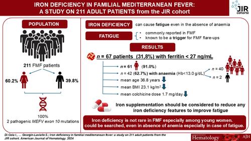
Overall, 211 FMF were included, mostly female (60.2%) and with a mean age of 41.34 [18–89] years. Out of 208 patients with the value of ferritin reported in our database, 67 (31.80%) had a serum ferritin level < 27 ng/mL and were defined as having iron deficiency. Among these, 61 (91.04%) were female and with a mean age of 36.81 [18–89] years. Among the 67 with iron deficiency, 42 (62.69%) displayed anemia (Hb < 13.0 g/dL), including 40 female and 2 males. Patients with iron deficiency showed lower values of Hb (p < 0.001), mean corpuscular volume (MCV) (p = 0.002), BMI (p = 0.044), weight (p < 0.001), daily colchicine dose (p = 0.015), and creatinine (p = 0.005) and they were younger than those without iron deficiency (p = 0.011). No differences were retrieved comparing these two groups regarding inflammatory markers. Particularly, 52.24% of FMF with ferritin < 27 showed normal inflammatory markers (Table S1). Among the 82 men, 6 showed a serum ferritin level < 27 ng/mL. These patients did not significantly differ in main features compared with those without iron deficiency (Table S2).
The difference between values of Hb and daily colchicine dose according to iron deficiency were also analyzed (Figure S1).
In our cohort, both univariate and multivariate regression models were built to evaluate the clinical risk profile of patients with ferritin < 27 ng/mL (Table 1). Based on univariate analyses, clinical relevance but also considering the number of patients characterized by ferritin < 27 ng/mL, we built two multivariate regression models. One model comprised age, female sex, CRP, SAA, BMI, and colchicine doses whereas the other included age, female sex, CRP, SAA, colchicine doses, and biological disease-modifying antirheumatic drugs (bDMARDs). Despite not significant univariate analysis, CRP and SAA were added in the models considering the possible influence of the inflammatory process on ferritin levels [6]. We added bDMARDs in a model considering that more severe patients were probably treated with these drugs. In the first model, age (OR: 0.97, 95% CI: 0.95–1.00, p = 0.041) and female sex (OR: 13.77, 95% CI: 4.96–38.18, p < 0.001) were significantly and independently associated with the presence of ferritin < 27 ng/mL. Similarly, in the other model, age (OR: 0.97, 95% CI: 0.95–1.00, p = 0.015), and female sex (OR: 16.15, 95% CI: 5.67–46.00, p < 0.001) significantly predicted ferritin < 27 ng/mL.
TABLE 1. Regression analyses evaluating the clinical risk profile of patients characterized by ferritin < 27 ng/mL.
Clinical variables
OR
SE
95% CI
p
Ferritin < 27 ng/mL
Univariate analysis
Age
0.98
0.009
0.95–1.00
0.063
Female sex
11.89
0.460
4.83–29.28
< 0.001
Hb
0.62
0.099
0.51–0.75
< 0.001
Hb < 13.0 g/dL
3.01
0.310
1.64–5.53
< 0.001
RBC
0.47
0.271
0.28–0.81
0.006
MCV
0.89
0.036
0.83–0.96
0.002
CRP
0.98
0.010
0.96–1.00
0.088
SAA
0.99
0.003
0.99–1.00
0.288
BMI
0.93
0.037
0.87–0.99
0.045
Daily colchicine dose
1.87
0.261
1.12–3.11
0.016
bDMARDs
0.74
0.377
0.36–1.56
0.431
Weight
0.96
0.013
0.93–0.98
< 0.001
Creatinine
0.94
0.014
0.91–0.97
< 0.001
Multivariate analysis
Age
0.97
0.013
0.95–1.00
0.041
Female sex
13.77
0.521
4.96–38.18
< 0.001
CRP
0.99
0.014
0.96–1.01
0.325
SAA
1.00
0.003
0.99–1.00
0.680
BMI
0.990
0.044
0.91–1.08
0.815
bDMARDs
1.56
0.325
0.83–2.96
0.170
Multivariate analysis
Age
0.97
0.013
0.95–1.00
0.015
Female sex
16.15
0.53
5.67–46.00
< 0.001
CRP
0.99
0.013
0.96–1.01
0.320
SAA
1.00
0.003
0.99–1.00
0.658
Daily colchicine dose
1.80
0.33
0.94–3.47
0.078
bDMARDs
1.67
0.52
0.61–4.63
0.322
Note: p < 0.05 was considered statistically significant.
Abbreviations: bDMARDs: biological disease-modifying antirheumatic drugs; BMI: body mass index; CRP: C reactive protein; Hb: hemoglobin; M: men; MCV: mean corpuscular volume; N: number of patients; SAA: serum amyloid A; SD: standard deviation; W: women.
Altogether, 31.8% of FMF displayed ferritin < 27 ng/mL and were mostly female and of younger age. Ferritin may be considered the mainstay for iron deficiency detection [3], which has been shown to be responsible for fatigue, a very frequent favoring factor in FMF flare-ups [2]. Although in absence of anemia, iron deficiency could be an additional factor in the onset of inflammatory flare-ups and may affect the patient's quality of life, although this aspect could not be fully studied retrospectively. Indeed, to clearly define an “iron deficiency” condition in FMF, also in absence of anemia, future studies are needed to assess other parameters of iron metabolism besides ferritin.
Based on clinical observations, and the predominance of young female, regardless of their inflammatory status, our first hypothesis is that ferritin < 27 ng/mL and associated iron deficiency, also in absence of anemia, may be secondary to excessive gynecological loss not entirely compensated by an increased intake of iron-rich foods. Due to the retrospective nature of this study and the fact that information on the characteristics of the patients' periods was not systematically collected, an exhaustive answer to this question could not be given, but when the affected women were interviewed, they most often reported heavy and long-lasting periods without taking contraceptive pill. It is also possible that young women eat less animal protein. Finally, digestive bleeding, potentially aggravated by frequent use of NSAIDs, should not be overlooked. Iron deficiency did not cause anemia in all patients, but it can cause fatigue, and eventually FMF attacks, suggesting the needing of iron supplementation.
Here, we showed that FMF patients with ferritin < 27 ng/mL had lower Hb and BMI and were significantly younger than others, without elevated inflammatory biomarkers. Interestingly, they required higher doses of colchicine, mainly 2 mg/day. The role of colchicine in decreasing iron intestinal absorption may be questioned. Our work does not allow us to answer this question, but not all patients with iron deficiency received the maximum daily colchicine dose.
In our opinion, it is important to correct iron deficiency in FMF, starting with treating the cause of the iron loss and correcting the oral intake if possible and tolerated. Oral iron combined with oral colchicine may cause digestive discomfort and transit disorders, leading patients to stop iron supplements. If oral supplementation is not possible, intravenous iron infusions can be offered. In addition to iron supplements, patients can be given a list of iron-rich foods. Finding the cause of the martial deficiency with a particular focus on excessive gynecological bleeding to reduce the frequency and duration of menstruation in women should be done. It is also important to look for associated gastric disorders, especially in this population with a probable excess risk, or Helicobacter pylori infection. Our study should be considered as “hypothesis generating” to provide the basis for further specific designed studies to better elucidate this issue. To the best of our knowledge, this would be the first analysis of value of ferritin in FMF. This could be of importance to evaluate the complicated mechanisms underlying the asthenia as trigger of an inflammatory disease. We underline that use the ferritin alone as the sole arbiter of iron deficiency has to be confirmed especially among patients with a monogenic autoinflammatory diseases such as FMF if they display inflammation.
This work highlights the importance of measuring ferritin levels in FMF, especially because this dosage is simple and available worldwide in routine care including some emerging countries where FMF is highly prevalent. In the presence of significant iron deficiency, iron supplementation should be done to correct it and reduce any iron deficiency signs such as asthenia and anemia that may lead to FMF attacks and also entertain an asthenia circle in FMF patients.
期刊介绍:
The American Journal of Hematology offers extensive coverage of experimental and clinical aspects of blood diseases in humans and animal models. The journal publishes original contributions in both non-malignant and malignant hematological diseases, encompassing clinical and basic studies in areas such as hemostasis, thrombosis, immunology, blood banking, and stem cell biology. Clinical translational reports highlighting innovative therapeutic approaches for the diagnosis and treatment of hematological diseases are actively encouraged.The American Journal of Hematology features regular original laboratory and clinical research articles, brief research reports, critical reviews, images in hematology, as well as letters and correspondence.