{"title":"上颌窦的体积分析和多探测器计算机断层扫描(MDCT)对各种鼻窦解剖变异的评估及其与慢性鼻窦炎的关系。","authors":"Aman Taneja, Ankur Malhotra, Shruti Chandak, Swasti Jain, Arpit Taneja, Deepti Arora, Swarna Laxmi, Aishwarya Pandey","doi":"10.25259/JCIS_124_2024","DOIUrl":null,"url":null,"abstract":"<p><strong>Objectives: </strong>The study aimed to evaluate the relationship between maxillary sinus volume and various sinonasal anatomical variants, as detected by multi-detector computed tomography, and their associations with chronic rhinosinusitis (CRS).</p><p><strong>Material and methods: </strong>A case-control study was conducted with 103 patients presenting with chronic sinonasal symptoms (cases) and 50 asymptomatic individuals (controls). A 128-slice computed tomography scanner was used to measure maxillary sinus volume and assess anatomical variants, such as a deviated nasal septum (DNS), concha bullosa (CB), and agger nasi cells. Exclusion criteria included previous sinonasal surgery, malignancy, craniofacial trauma, and lack of consent. Statistical analysis was performed using <i>t</i>-tests for continuous variables and Chi-square tests for categorical data. Receiver operating characteristic curve analysis was utilized to determine a DNS angle cutoff for predicting CRS.</p><p><strong>Results: </strong>Anatomical variants were significantly more frequent in cases than in controls. The left-sided DNS was predominant in cases, while the right-sided DNS was more common in controls. The mean DNS deviation angle was notably larger in cases (10.84° ± 7.87) than in controls (5.55° ± 5.02). Maxillary sinus volume was significantly smaller in cases (9.69 cc on the left side and 10.23 cc on the right side) compared to controls (18.57 cc and 18.46 cc, respectively), with female patients exhibiting smaller volumes than males. Agger nasi cells were detected in 51.5% of cases versus 8.0% of controls. A strong association was found between CB and contralateral DNS. The optimal DNS deviation angle cutoff for predicting CRS was identified as 12.7°.</p><p><strong>Conclusion: </strong>This study shows that CRS is linked to smaller maxillary sinus volumes, with males having larger sinus volumes than females. A DNS and larger deviation angles were associated with a higher risk of sinus inflammation, with angles over 12.7° predicting the onset of the condition. The presence of CB and agger nasi cells also contributed to the development of CRS.</p>","PeriodicalId":15512,"journal":{"name":"Journal of Clinical Imaging Science","volume":"14 ","pages":"44"},"PeriodicalIF":1.3000,"publicationDate":"2024-11-18","publicationTypes":"Journal Article","fieldsOfStudy":null,"isOpenAccess":false,"openAccessPdf":"https://www.ncbi.nlm.nih.gov/pmc/articles/PMC11618756/pdf/","citationCount":"0","resultStr":"{\"title\":\"Volumetric analysis of maxillary sinus and assessment of various sinonasal anatomic variants on multi-detector computed tomography (MDCT) and their association with chronic rhinosinusitis.\",\"authors\":\"Aman Taneja, Ankur Malhotra, Shruti Chandak, Swasti Jain, Arpit Taneja, Deepti Arora, Swarna Laxmi, Aishwarya Pandey\",\"doi\":\"10.25259/JCIS_124_2024\",\"DOIUrl\":null,\"url\":null,\"abstract\":\"<p><strong>Objectives: </strong>The study aimed to evaluate the relationship between maxillary sinus volume and various sinonasal anatomical variants, as detected by multi-detector computed tomography, and their associations with chronic rhinosinusitis (CRS).</p><p><strong>Material and methods: </strong>A case-control study was conducted with 103 patients presenting with chronic sinonasal symptoms (cases) and 50 asymptomatic individuals (controls). A 128-slice computed tomography scanner was used to measure maxillary sinus volume and assess anatomical variants, such as a deviated nasal septum (DNS), concha bullosa (CB), and agger nasi cells. Exclusion criteria included previous sinonasal surgery, malignancy, craniofacial trauma, and lack of consent. Statistical analysis was performed using <i>t</i>-tests for continuous variables and Chi-square tests for categorical data. Receiver operating characteristic curve analysis was utilized to determine a DNS angle cutoff for predicting CRS.</p><p><strong>Results: </strong>Anatomical variants were significantly more frequent in cases than in controls. The left-sided DNS was predominant in cases, while the right-sided DNS was more common in controls. The mean DNS deviation angle was notably larger in cases (10.84° ± 7.87) than in controls (5.55° ± 5.02). Maxillary sinus volume was significantly smaller in cases (9.69 cc on the left side and 10.23 cc on the right side) compared to controls (18.57 cc and 18.46 cc, respectively), with female patients exhibiting smaller volumes than males. Agger nasi cells were detected in 51.5% of cases versus 8.0% of controls. A strong association was found between CB and contralateral DNS. The optimal DNS deviation angle cutoff for predicting CRS was identified as 12.7°.</p><p><strong>Conclusion: </strong>This study shows that CRS is linked to smaller maxillary sinus volumes, with males having larger sinus volumes than females. A DNS and larger deviation angles were associated with a higher risk of sinus inflammation, with angles over 12.7° predicting the onset of the condition. The presence of CB and agger nasi cells also contributed to the development of CRS.</p>\",\"PeriodicalId\":15512,\"journal\":{\"name\":\"Journal of Clinical Imaging Science\",\"volume\":\"14 \",\"pages\":\"44\"},\"PeriodicalIF\":1.3000,\"publicationDate\":\"2024-11-18\",\"publicationTypes\":\"Journal Article\",\"fieldsOfStudy\":null,\"isOpenAccess\":false,\"openAccessPdf\":\"https://www.ncbi.nlm.nih.gov/pmc/articles/PMC11618756/pdf/\",\"citationCount\":\"0\",\"resultStr\":null,\"platform\":\"Semanticscholar\",\"paperid\":null,\"PeriodicalName\":\"Journal of Clinical Imaging Science\",\"FirstCategoryId\":\"1085\",\"ListUrlMain\":\"https://doi.org/10.25259/JCIS_124_2024\",\"RegionNum\":0,\"RegionCategory\":null,\"ArticlePicture\":[],\"TitleCN\":null,\"AbstractTextCN\":null,\"PMCID\":null,\"EPubDate\":\"2024/1/1 0:00:00\",\"PubModel\":\"eCollection\",\"JCR\":\"Q3\",\"JCRName\":\"RADIOLOGY, NUCLEAR MEDICINE & MEDICAL IMAGING\",\"Score\":null,\"Total\":0}","platform":"Semanticscholar","paperid":null,"PeriodicalName":"Journal of Clinical Imaging Science","FirstCategoryId":"1085","ListUrlMain":"https://doi.org/10.25259/JCIS_124_2024","RegionNum":0,"RegionCategory":null,"ArticlePicture":[],"TitleCN":null,"AbstractTextCN":null,"PMCID":null,"EPubDate":"2024/1/1 0:00:00","PubModel":"eCollection","JCR":"Q3","JCRName":"RADIOLOGY, NUCLEAR MEDICINE & MEDICAL IMAGING","Score":null,"Total":0}
Volumetric analysis of maxillary sinus and assessment of various sinonasal anatomic variants on multi-detector computed tomography (MDCT) and their association with chronic rhinosinusitis.
Objectives: The study aimed to evaluate the relationship between maxillary sinus volume and various sinonasal anatomical variants, as detected by multi-detector computed tomography, and their associations with chronic rhinosinusitis (CRS).
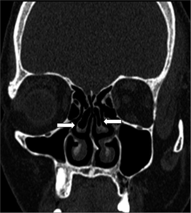
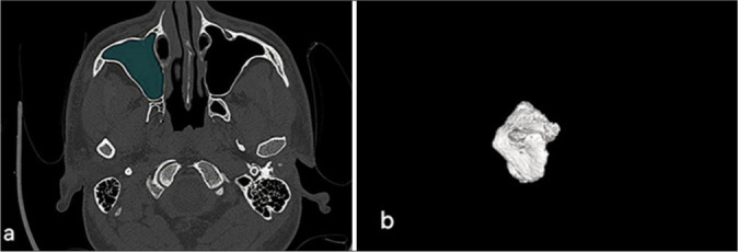
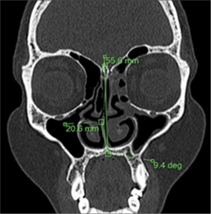
Material and methods: A case-control study was conducted with 103 patients presenting with chronic sinonasal symptoms (cases) and 50 asymptomatic individuals (controls). A 128-slice computed tomography scanner was used to measure maxillary sinus volume and assess anatomical variants, such as a deviated nasal septum (DNS), concha bullosa (CB), and agger nasi cells. Exclusion criteria included previous sinonasal surgery, malignancy, craniofacial trauma, and lack of consent. Statistical analysis was performed using t-tests for continuous variables and Chi-square tests for categorical data. Receiver operating characteristic curve analysis was utilized to determine a DNS angle cutoff for predicting CRS.
Results: Anatomical variants were significantly more frequent in cases than in controls. The left-sided DNS was predominant in cases, while the right-sided DNS was more common in controls. The mean DNS deviation angle was notably larger in cases (10.84° ± 7.87) than in controls (5.55° ± 5.02). Maxillary sinus volume was significantly smaller in cases (9.69 cc on the left side and 10.23 cc on the right side) compared to controls (18.57 cc and 18.46 cc, respectively), with female patients exhibiting smaller volumes than males. Agger nasi cells were detected in 51.5% of cases versus 8.0% of controls. A strong association was found between CB and contralateral DNS. The optimal DNS deviation angle cutoff for predicting CRS was identified as 12.7°.
Conclusion: This study shows that CRS is linked to smaller maxillary sinus volumes, with males having larger sinus volumes than females. A DNS and larger deviation angles were associated with a higher risk of sinus inflammation, with angles over 12.7° predicting the onset of the condition. The presence of CB and agger nasi cells also contributed to the development of CRS.
期刊介绍:
The Journal of Clinical Imaging Science (JCIS) is an open access peer-reviewed journal committed to publishing high-quality articles in the field of Imaging Science. The journal aims to present Imaging Science and relevant clinical information in an understandable and useful format. The journal is owned and published by the Scientific Scholar. Audience Our audience includes Radiologists, Researchers, Clinicians, medical professionals and students. Review process JCIS has a highly rigorous peer-review process that makes sure that manuscripts are scientifically accurate, relevant, novel and important. Authors disclose all conflicts, affiliations and financial associations such that the published content is not biased.

 求助内容:
求助内容: 应助结果提醒方式:


