{"title":"增强磁共振图像和下游分割,配准和诊断任务的基础模型","authors":"Yue Sun, Limei Wang, Gang Li, Weili Lin, Li Wang","doi":"10.1038/s41551-024-01283-7","DOIUrl":null,"url":null,"abstract":"<p>In structural magnetic resonance (MR) imaging, motion artefacts, low resolution, imaging noise and variability in acquisition protocols frequently degrade image quality and confound downstream analyses. Here we report a foundation model for the motion correction, resolution enhancement, denoising and harmonization of MR images. Specifically, we trained a tissue-classification neural network to predict tissue labels, which are then leveraged by a ‘tissue-aware’ enhancement network to generate high-quality MR images. We validated the model’s effectiveness on a large and diverse dataset comprising 2,448 deliberately corrupted images and 10,963 images spanning a wide age range (from foetuses to elderly individuals) acquired using a variety of clinical scanners across 19 public datasets. The model consistently outperformed state-of-the-art algorithms in improving the quality of MR images, handling pathological brains with multiple sclerosis or gliomas, generating 7-T-like images from 3 T scans and harmonizing images acquired from different scanners. The high-quality, high-resolution and harmonized images generated by the model can be used to enhance the performance of models for tissue segmentation, registration, diagnosis and other downstream tasks.</p>","PeriodicalId":19063,"journal":{"name":"Nature Biomedical Engineering","volume":"53 1","pages":""},"PeriodicalIF":26.8000,"publicationDate":"2024-12-05","publicationTypes":"Journal Article","fieldsOfStudy":null,"isOpenAccess":false,"openAccessPdf":"","citationCount":"0","resultStr":"{\"title\":\"A foundation model for enhancing magnetic resonance images and downstream segmentation, registration and diagnostic tasks\",\"authors\":\"Yue Sun, Limei Wang, Gang Li, Weili Lin, Li Wang\",\"doi\":\"10.1038/s41551-024-01283-7\",\"DOIUrl\":null,\"url\":null,\"abstract\":\"<p>In structural magnetic resonance (MR) imaging, motion artefacts, low resolution, imaging noise and variability in acquisition protocols frequently degrade image quality and confound downstream analyses. Here we report a foundation model for the motion correction, resolution enhancement, denoising and harmonization of MR images. Specifically, we trained a tissue-classification neural network to predict tissue labels, which are then leveraged by a ‘tissue-aware’ enhancement network to generate high-quality MR images. We validated the model’s effectiveness on a large and diverse dataset comprising 2,448 deliberately corrupted images and 10,963 images spanning a wide age range (from foetuses to elderly individuals) acquired using a variety of clinical scanners across 19 public datasets. The model consistently outperformed state-of-the-art algorithms in improving the quality of MR images, handling pathological brains with multiple sclerosis or gliomas, generating 7-T-like images from 3 T scans and harmonizing images acquired from different scanners. The high-quality, high-resolution and harmonized images generated by the model can be used to enhance the performance of models for tissue segmentation, registration, diagnosis and other downstream tasks.</p>\",\"PeriodicalId\":19063,\"journal\":{\"name\":\"Nature Biomedical Engineering\",\"volume\":\"53 1\",\"pages\":\"\"},\"PeriodicalIF\":26.8000,\"publicationDate\":\"2024-12-05\",\"publicationTypes\":\"Journal Article\",\"fieldsOfStudy\":null,\"isOpenAccess\":false,\"openAccessPdf\":\"\",\"citationCount\":\"0\",\"resultStr\":null,\"platform\":\"Semanticscholar\",\"paperid\":null,\"PeriodicalName\":\"Nature Biomedical Engineering\",\"FirstCategoryId\":\"5\",\"ListUrlMain\":\"https://doi.org/10.1038/s41551-024-01283-7\",\"RegionNum\":1,\"RegionCategory\":\"医学\",\"ArticlePicture\":[],\"TitleCN\":null,\"AbstractTextCN\":null,\"PMCID\":null,\"EPubDate\":\"\",\"PubModel\":\"\",\"JCR\":\"Q1\",\"JCRName\":\"ENGINEERING, BIOMEDICAL\",\"Score\":null,\"Total\":0}","platform":"Semanticscholar","paperid":null,"PeriodicalName":"Nature Biomedical Engineering","FirstCategoryId":"5","ListUrlMain":"https://doi.org/10.1038/s41551-024-01283-7","RegionNum":1,"RegionCategory":"医学","ArticlePicture":[],"TitleCN":null,"AbstractTextCN":null,"PMCID":null,"EPubDate":"","PubModel":"","JCR":"Q1","JCRName":"ENGINEERING, BIOMEDICAL","Score":null,"Total":0}
A foundation model for enhancing magnetic resonance images and downstream segmentation, registration and diagnostic tasks
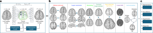
In structural magnetic resonance (MR) imaging, motion artefacts, low resolution, imaging noise and variability in acquisition protocols frequently degrade image quality and confound downstream analyses. Here we report a foundation model for the motion correction, resolution enhancement, denoising and harmonization of MR images. Specifically, we trained a tissue-classification neural network to predict tissue labels, which are then leveraged by a ‘tissue-aware’ enhancement network to generate high-quality MR images. We validated the model’s effectiveness on a large and diverse dataset comprising 2,448 deliberately corrupted images and 10,963 images spanning a wide age range (from foetuses to elderly individuals) acquired using a variety of clinical scanners across 19 public datasets. The model consistently outperformed state-of-the-art algorithms in improving the quality of MR images, handling pathological brains with multiple sclerosis or gliomas, generating 7-T-like images from 3 T scans and harmonizing images acquired from different scanners. The high-quality, high-resolution and harmonized images generated by the model can be used to enhance the performance of models for tissue segmentation, registration, diagnosis and other downstream tasks.
期刊介绍:
Nature Biomedical Engineering is an online-only monthly journal that was launched in January 2017. It aims to publish original research, reviews, and commentary focusing on applied biomedicine and health technology. The journal targets a diverse audience, including life scientists who are involved in developing experimental or computational systems and methods to enhance our understanding of human physiology. It also covers biomedical researchers and engineers who are engaged in designing or optimizing therapies, assays, devices, or procedures for diagnosing or treating diseases. Additionally, clinicians, who make use of research outputs to evaluate patient health or administer therapy in various clinical settings and healthcare contexts, are also part of the target audience.
 求助内容:
求助内容: 应助结果提醒方式:


