Giorgio Tamborrini, Raphael Micheroli, Vincenzo Ricci, Marco Becciolini, Mario Garcia-Pompermayer, Andres Serrano Belmar Gonzalo, Magdalena Müller-Gerbl, Felix Margenfeld
{"title":"推进高分辨率肌肉骨骼超声:一种组织学和解剖学驱动的方法,用于增强肩部成像。第一部分:后冠状肩。","authors":"Giorgio Tamborrini, Raphael Micheroli, Vincenzo Ricci, Marco Becciolini, Mario Garcia-Pompermayer, Andres Serrano Belmar Gonzalo, Magdalena Müller-Gerbl, Felix Margenfeld","doi":"10.15557/jou.2024.0026","DOIUrl":null,"url":null,"abstract":"<p><p>Ultrasound is a reliable imaging modality for diagnosing and assessing musculoskeletal disorders. Recent advancements in ultrasound technology have substantially improved image resolution, enabling the visualization of anatomic structures on a near-microscopic level. However, existing guidelines for standardized shoulder ultrasound primarily rely on earlier machine models and settings that may not harness the full potential of these high-resolution imaging capabilities. This article provides a simple and systematic guide to high-resolution sonography of the shoulder using anatomical and histological images from cadavers for comparison. International standard techniques are considered, and images were obtained using modern equipment. This two-article series systematically shows the examination and normal findings, presenting first the posterior, then frontal, then further anterior, followed by lateral and, optionally, the axillary examination. In this article, the focus is on the posterior and coronal shoulder.</p>","PeriodicalId":45612,"journal":{"name":"Journal of Ultrasonography","volume":"24 98","pages":"1-9"},"PeriodicalIF":1.5000,"publicationDate":"2024-11-30","publicationTypes":"Journal Article","fieldsOfStudy":null,"isOpenAccess":false,"openAccessPdf":"https://www.ncbi.nlm.nih.gov/pmc/articles/PMC11608069/pdf/","citationCount":"0","resultStr":"{\"title\":\"Advancing high-resolution musculoskeletal ultrasound: A histology- and anatomy-driven approach for enhanced shoulder imaging. Part I: Posterior and coronal shoulder.\",\"authors\":\"Giorgio Tamborrini, Raphael Micheroli, Vincenzo Ricci, Marco Becciolini, Mario Garcia-Pompermayer, Andres Serrano Belmar Gonzalo, Magdalena Müller-Gerbl, Felix Margenfeld\",\"doi\":\"10.15557/jou.2024.0026\",\"DOIUrl\":null,\"url\":null,\"abstract\":\"<p><p>Ultrasound is a reliable imaging modality for diagnosing and assessing musculoskeletal disorders. Recent advancements in ultrasound technology have substantially improved image resolution, enabling the visualization of anatomic structures on a near-microscopic level. However, existing guidelines for standardized shoulder ultrasound primarily rely on earlier machine models and settings that may not harness the full potential of these high-resolution imaging capabilities. This article provides a simple and systematic guide to high-resolution sonography of the shoulder using anatomical and histological images from cadavers for comparison. International standard techniques are considered, and images were obtained using modern equipment. This two-article series systematically shows the examination and normal findings, presenting first the posterior, then frontal, then further anterior, followed by lateral and, optionally, the axillary examination. In this article, the focus is on the posterior and coronal shoulder.</p>\",\"PeriodicalId\":45612,\"journal\":{\"name\":\"Journal of Ultrasonography\",\"volume\":\"24 98\",\"pages\":\"1-9\"},\"PeriodicalIF\":1.5000,\"publicationDate\":\"2024-11-30\",\"publicationTypes\":\"Journal Article\",\"fieldsOfStudy\":null,\"isOpenAccess\":false,\"openAccessPdf\":\"https://www.ncbi.nlm.nih.gov/pmc/articles/PMC11608069/pdf/\",\"citationCount\":\"0\",\"resultStr\":null,\"platform\":\"Semanticscholar\",\"paperid\":null,\"PeriodicalName\":\"Journal of Ultrasonography\",\"FirstCategoryId\":\"1085\",\"ListUrlMain\":\"https://doi.org/10.15557/jou.2024.0026\",\"RegionNum\":0,\"RegionCategory\":null,\"ArticlePicture\":[],\"TitleCN\":null,\"AbstractTextCN\":null,\"PMCID\":null,\"EPubDate\":\"2024/12/1 0:00:00\",\"PubModel\":\"eCollection\",\"JCR\":\"Q3\",\"JCRName\":\"RADIOLOGY, NUCLEAR MEDICINE & MEDICAL IMAGING\",\"Score\":null,\"Total\":0}","platform":"Semanticscholar","paperid":null,"PeriodicalName":"Journal of Ultrasonography","FirstCategoryId":"1085","ListUrlMain":"https://doi.org/10.15557/jou.2024.0026","RegionNum":0,"RegionCategory":null,"ArticlePicture":[],"TitleCN":null,"AbstractTextCN":null,"PMCID":null,"EPubDate":"2024/12/1 0:00:00","PubModel":"eCollection","JCR":"Q3","JCRName":"RADIOLOGY, NUCLEAR MEDICINE & MEDICAL IMAGING","Score":null,"Total":0}
Advancing high-resolution musculoskeletal ultrasound: A histology- and anatomy-driven approach for enhanced shoulder imaging. Part I: Posterior and coronal shoulder.
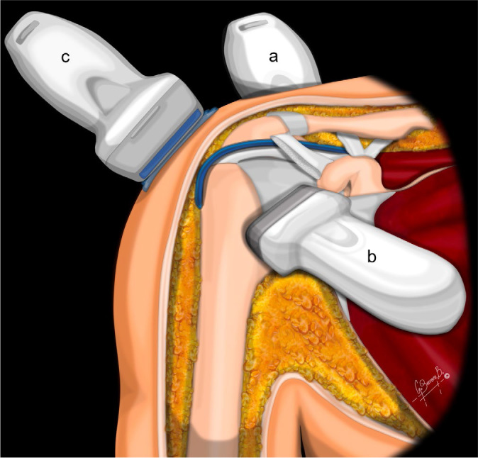
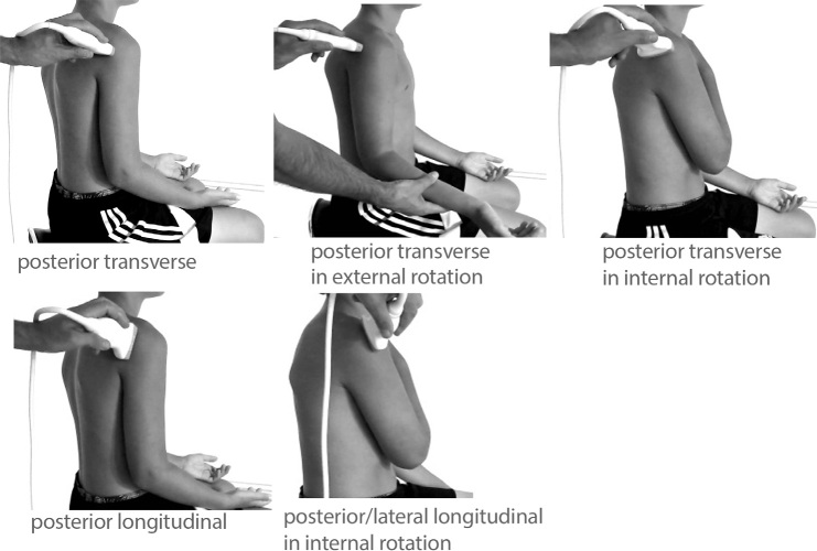
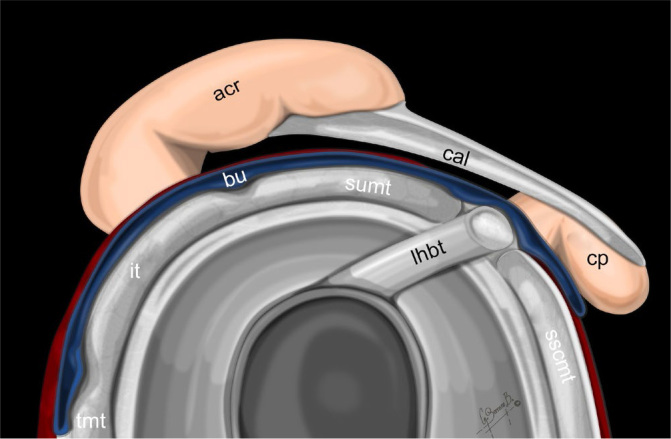
Ultrasound is a reliable imaging modality for diagnosing and assessing musculoskeletal disorders. Recent advancements in ultrasound technology have substantially improved image resolution, enabling the visualization of anatomic structures on a near-microscopic level. However, existing guidelines for standardized shoulder ultrasound primarily rely on earlier machine models and settings that may not harness the full potential of these high-resolution imaging capabilities. This article provides a simple and systematic guide to high-resolution sonography of the shoulder using anatomical and histological images from cadavers for comparison. International standard techniques are considered, and images were obtained using modern equipment. This two-article series systematically shows the examination and normal findings, presenting first the posterior, then frontal, then further anterior, followed by lateral and, optionally, the axillary examination. In this article, the focus is on the posterior and coronal shoulder.

 求助内容:
求助内容: 应助结果提醒方式:


