Hyeseon Lee, Sijoon Lee, Seung Yun Yang, Dong Hwan Kim, Mahnjeong Ha, Kyoung Hyup Nam
{"title":"基于透明质酸的可生物降解光固化密封剂的降解模式:大鼠颅骨切除术模型的一系列磁共振成像观察研究。","authors":"Hyeseon Lee, Sijoon Lee, Seung Yun Yang, Dong Hwan Kim, Mahnjeong Ha, Kyoung Hyup Nam","doi":"10.3340/jkns.2024.0138","DOIUrl":null,"url":null,"abstract":"<p><strong>Objective: </strong>The aim of this study is evaluating in vivo degradation of photocrosslinkable hyaluronic acid (HA)-based dural sealant (HA photosealant) using magnetic resonance imaging (MRI) and histopathological analysis to assess its biodegradability and effectiveness in preventing cerebrospinal fluid (CSF) leakage.</p><p><strong>Methods: </strong>HA photosealants were applied to the incised dura in a rat craniectomy and durotomy. The HA photosealant quickly sealed the wound upon low-energy visible light exposure (405 nm, <5 seconds, < 1 J/cm2). The degradation of HA photosealants was tracked through serial MRI scans at 1, 2, and 4 weeks post-application. The remaining area of HA photosealants on the dura was measured using image processing program for volumetric analysis. Additionally, histopathological analyses were performed to evaluate the biocompatibility and effectiveness of the dural repair.</p><p><strong>Results: </strong>The MRI and histopathological analyses showed that the HA photosealant achieved progressive degradation while successfully preventing CSF leakage without any adverse tissue reactions. The residual area of HA photosealants measured at 2 weeks ranged from 41.35% to 94.88%, with an average of 66.57%. At 4 weeks, a more distinct degradation pattern was observed compared to 2 weeks, showing a residual area of 10.28% to 56.11%. The HA photosealant maintained structural integrity until dural regeneration was completed.</p><p><strong>Conclusion: </strong>HA photosealant showed gradual degradation in vivo while maintaining mechanical strength until the dura was repaired for preventing CSF leakage without inflammation and toxicity. HA photosealant has great potentials for clinical application for dural repair with biodegradable properties and biocompatibility.</p>","PeriodicalId":16283,"journal":{"name":"Journal of Korean Neurosurgical Society","volume":" ","pages":"375-382"},"PeriodicalIF":1.7000,"publicationDate":"2025-07-01","publicationTypes":"Journal Article","fieldsOfStudy":null,"isOpenAccess":false,"openAccessPdf":"https://www.ncbi.nlm.nih.gov/pmc/articles/PMC12237541/pdf/","citationCount":"0","resultStr":"{\"title\":\"Degradation Pattern of a Biodegradable and Photocurable Sealants Based on Hyaluronic Acid : A Serial Magnetic Resonance Imaging Observational Study in Rat Craniectomy Model.\",\"authors\":\"Hyeseon Lee, Sijoon Lee, Seung Yun Yang, Dong Hwan Kim, Mahnjeong Ha, Kyoung Hyup Nam\",\"doi\":\"10.3340/jkns.2024.0138\",\"DOIUrl\":null,\"url\":null,\"abstract\":\"<p><strong>Objective: </strong>The aim of this study is evaluating in vivo degradation of photocrosslinkable hyaluronic acid (HA)-based dural sealant (HA photosealant) using magnetic resonance imaging (MRI) and histopathological analysis to assess its biodegradability and effectiveness in preventing cerebrospinal fluid (CSF) leakage.</p><p><strong>Methods: </strong>HA photosealants were applied to the incised dura in a rat craniectomy and durotomy. The HA photosealant quickly sealed the wound upon low-energy visible light exposure (405 nm, <5 seconds, < 1 J/cm2). The degradation of HA photosealants was tracked through serial MRI scans at 1, 2, and 4 weeks post-application. The remaining area of HA photosealants on the dura was measured using image processing program for volumetric analysis. Additionally, histopathological analyses were performed to evaluate the biocompatibility and effectiveness of the dural repair.</p><p><strong>Results: </strong>The MRI and histopathological analyses showed that the HA photosealant achieved progressive degradation while successfully preventing CSF leakage without any adverse tissue reactions. The residual area of HA photosealants measured at 2 weeks ranged from 41.35% to 94.88%, with an average of 66.57%. At 4 weeks, a more distinct degradation pattern was observed compared to 2 weeks, showing a residual area of 10.28% to 56.11%. The HA photosealant maintained structural integrity until dural regeneration was completed.</p><p><strong>Conclusion: </strong>HA photosealant showed gradual degradation in vivo while maintaining mechanical strength until the dura was repaired for preventing CSF leakage without inflammation and toxicity. HA photosealant has great potentials for clinical application for dural repair with biodegradable properties and biocompatibility.</p>\",\"PeriodicalId\":16283,\"journal\":{\"name\":\"Journal of Korean Neurosurgical Society\",\"volume\":\" \",\"pages\":\"375-382\"},\"PeriodicalIF\":1.7000,\"publicationDate\":\"2025-07-01\",\"publicationTypes\":\"Journal Article\",\"fieldsOfStudy\":null,\"isOpenAccess\":false,\"openAccessPdf\":\"https://www.ncbi.nlm.nih.gov/pmc/articles/PMC12237541/pdf/\",\"citationCount\":\"0\",\"resultStr\":null,\"platform\":\"Semanticscholar\",\"paperid\":null,\"PeriodicalName\":\"Journal of Korean Neurosurgical Society\",\"FirstCategoryId\":\"3\",\"ListUrlMain\":\"https://doi.org/10.3340/jkns.2024.0138\",\"RegionNum\":4,\"RegionCategory\":\"医学\",\"ArticlePicture\":[],\"TitleCN\":null,\"AbstractTextCN\":null,\"PMCID\":null,\"EPubDate\":\"2024/12/3 0:00:00\",\"PubModel\":\"Epub\",\"JCR\":\"Q4\",\"JCRName\":\"CLINICAL NEUROLOGY\",\"Score\":null,\"Total\":0}","platform":"Semanticscholar","paperid":null,"PeriodicalName":"Journal of Korean Neurosurgical Society","FirstCategoryId":"3","ListUrlMain":"https://doi.org/10.3340/jkns.2024.0138","RegionNum":4,"RegionCategory":"医学","ArticlePicture":[],"TitleCN":null,"AbstractTextCN":null,"PMCID":null,"EPubDate":"2024/12/3 0:00:00","PubModel":"Epub","JCR":"Q4","JCRName":"CLINICAL NEUROLOGY","Score":null,"Total":0}
Degradation Pattern of a Biodegradable and Photocurable Sealants Based on Hyaluronic Acid : A Serial Magnetic Resonance Imaging Observational Study in Rat Craniectomy Model.
Objective: The aim of this study is evaluating in vivo degradation of photocrosslinkable hyaluronic acid (HA)-based dural sealant (HA photosealant) using magnetic resonance imaging (MRI) and histopathological analysis to assess its biodegradability and effectiveness in preventing cerebrospinal fluid (CSF) leakage.
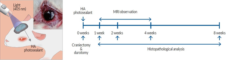
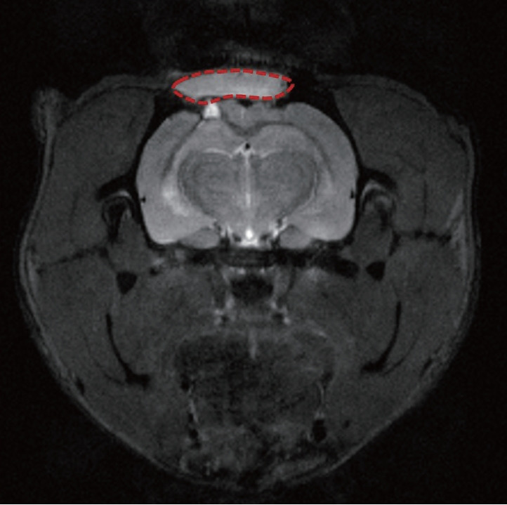
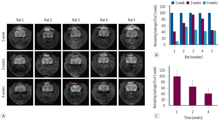
Methods: HA photosealants were applied to the incised dura in a rat craniectomy and durotomy. The HA photosealant quickly sealed the wound upon low-energy visible light exposure (405 nm, <5 seconds, < 1 J/cm2). The degradation of HA photosealants was tracked through serial MRI scans at 1, 2, and 4 weeks post-application. The remaining area of HA photosealants on the dura was measured using image processing program for volumetric analysis. Additionally, histopathological analyses were performed to evaluate the biocompatibility and effectiveness of the dural repair.
Results: The MRI and histopathological analyses showed that the HA photosealant achieved progressive degradation while successfully preventing CSF leakage without any adverse tissue reactions. The residual area of HA photosealants measured at 2 weeks ranged from 41.35% to 94.88%, with an average of 66.57%. At 4 weeks, a more distinct degradation pattern was observed compared to 2 weeks, showing a residual area of 10.28% to 56.11%. The HA photosealant maintained structural integrity until dural regeneration was completed.
Conclusion: HA photosealant showed gradual degradation in vivo while maintaining mechanical strength until the dura was repaired for preventing CSF leakage without inflammation and toxicity. HA photosealant has great potentials for clinical application for dural repair with biodegradable properties and biocompatibility.
期刊介绍:
The Journal of Korean Neurosurgical Society (J Korean Neurosurg Soc) is the official journal of the Korean Neurosurgical Society, and published bimonthly (1st day of January, March, May, July, September, and November). It launched in October 31, 1972 with Volume 1 and Number 1. J Korean Neurosurg Soc aims to allow neurosurgeons from around the world to enrich their knowledge of patient management, education, and clinical or experimental research, and hence their professionalism. This journal publishes Laboratory Investigations, Clinical Articles, Review Articles, Case Reports, Technical Notes, and Letters to the Editor. Our field of interest involves clinical neurosurgery (cerebrovascular disease, neuro-oncology, skull base neurosurgery, spine, pediatric neurosurgery, functional neurosurgery, epilepsy, neuro-trauma, and peripheral nerve disease) and laboratory work in neuroscience.
 求助内容:
求助内容: 应助结果提醒方式:


