S Gadallah, A Sharshar, M Fadel, E Mahran, A Hammad
{"title":"埃及驴大炮区掌/足底方面的肌腱和韧带的超声特征。","authors":"S Gadallah, A Sharshar, M Fadel, E Mahran, A Hammad","doi":"10.22099/IJVR.2024.47480.6859","DOIUrl":null,"url":null,"abstract":"<p><strong>Background: </strong>There is a scarcity of data regarding the ultrasonographic characterization of tendons and ligaments in the distal limbs of donkeys.</p><p><strong>Aims: </strong>To determine ultrasonographic characteristics of normal tendons and ligaments at the palmar/plantar aspect of the cannon region in Egyptian donkeys.</p><p><strong>Methods: </strong>B-mode ultrasonography was conducted for the proposed tendons and ligaments in 12 clinically normal donkeys. Targeted structures were examined using transverse and longitudinal scans and evaluated in shape, echogenicity, echogenic pattern, fiber alignment pattern, and cross-sectional area (CSA).</p><p><strong>Results: </strong>Using transverse scan, the sonographic shapes of tendons and ligaments of metacarpal and metatarsal regions were determined. Upon examining different levels of each region, specific ligaments, not tendons, were present only at the higher levels. The echogenicity of ligaments and tendons was either similar or variable across different levels. All tendons and ligaments displayed homogeneous echogenicity except for the suspensory ligament. In the longitudinal scan, tendons maintained linear and parallel fiber alignment along examination levels. Conversely, ligaments displayed mostly inconstant fiber patterns (linear/crimp). There was a statistically significant (P<0.05) difference in CSA of tendons and ligaments between certain levels within metacarpal and metatarsal regions. Upon comparing metacarpal and metatarsal regions, there were distinct variations in some ultrasonographic characteristics of the same tendons and ligaments.</p><p><strong>Conclusion: </strong>This study established the ultrasonographic features of normal tendons and ligaments at the palmar/plantar aspect of the cannon region in donkeys (<i>Equus asinus</i>). These ultrasonographic data can be a reference guide when cannon region lameness is suspected.</p>","PeriodicalId":14629,"journal":{"name":"Iranian journal of veterinary research","volume":"25 2","pages":"143-155"},"PeriodicalIF":1.0000,"publicationDate":"2024-01-01","publicationTypes":"Journal Article","fieldsOfStudy":null,"isOpenAccess":false,"openAccessPdf":"https://www.ncbi.nlm.nih.gov/pmc/articles/PMC11608532/pdf/","citationCount":"0","resultStr":"{\"title\":\"Ultrasonographic characterization of tendons and ligaments of palmar/plantar aspect of the cannon region in Egyptian donkeys.\",\"authors\":\"S Gadallah, A Sharshar, M Fadel, E Mahran, A Hammad\",\"doi\":\"10.22099/IJVR.2024.47480.6859\",\"DOIUrl\":null,\"url\":null,\"abstract\":\"<p><strong>Background: </strong>There is a scarcity of data regarding the ultrasonographic characterization of tendons and ligaments in the distal limbs of donkeys.</p><p><strong>Aims: </strong>To determine ultrasonographic characteristics of normal tendons and ligaments at the palmar/plantar aspect of the cannon region in Egyptian donkeys.</p><p><strong>Methods: </strong>B-mode ultrasonography was conducted for the proposed tendons and ligaments in 12 clinically normal donkeys. Targeted structures were examined using transverse and longitudinal scans and evaluated in shape, echogenicity, echogenic pattern, fiber alignment pattern, and cross-sectional area (CSA).</p><p><strong>Results: </strong>Using transverse scan, the sonographic shapes of tendons and ligaments of metacarpal and metatarsal regions were determined. Upon examining different levels of each region, specific ligaments, not tendons, were present only at the higher levels. The echogenicity of ligaments and tendons was either similar or variable across different levels. All tendons and ligaments displayed homogeneous echogenicity except for the suspensory ligament. In the longitudinal scan, tendons maintained linear and parallel fiber alignment along examination levels. Conversely, ligaments displayed mostly inconstant fiber patterns (linear/crimp). There was a statistically significant (P<0.05) difference in CSA of tendons and ligaments between certain levels within metacarpal and metatarsal regions. Upon comparing metacarpal and metatarsal regions, there were distinct variations in some ultrasonographic characteristics of the same tendons and ligaments.</p><p><strong>Conclusion: </strong>This study established the ultrasonographic features of normal tendons and ligaments at the palmar/plantar aspect of the cannon region in donkeys (<i>Equus asinus</i>). These ultrasonographic data can be a reference guide when cannon region lameness is suspected.</p>\",\"PeriodicalId\":14629,\"journal\":{\"name\":\"Iranian journal of veterinary research\",\"volume\":\"25 2\",\"pages\":\"143-155\"},\"PeriodicalIF\":1.0000,\"publicationDate\":\"2024-01-01\",\"publicationTypes\":\"Journal Article\",\"fieldsOfStudy\":null,\"isOpenAccess\":false,\"openAccessPdf\":\"https://www.ncbi.nlm.nih.gov/pmc/articles/PMC11608532/pdf/\",\"citationCount\":\"0\",\"resultStr\":null,\"platform\":\"Semanticscholar\",\"paperid\":null,\"PeriodicalName\":\"Iranian journal of veterinary research\",\"FirstCategoryId\":\"97\",\"ListUrlMain\":\"https://doi.org/10.22099/IJVR.2024.47480.6859\",\"RegionNum\":4,\"RegionCategory\":\"农林科学\",\"ArticlePicture\":[],\"TitleCN\":null,\"AbstractTextCN\":null,\"PMCID\":null,\"EPubDate\":\"\",\"PubModel\":\"\",\"JCR\":\"Q3\",\"JCRName\":\"VETERINARY SCIENCES\",\"Score\":null,\"Total\":0}","platform":"Semanticscholar","paperid":null,"PeriodicalName":"Iranian journal of veterinary research","FirstCategoryId":"97","ListUrlMain":"https://doi.org/10.22099/IJVR.2024.47480.6859","RegionNum":4,"RegionCategory":"农林科学","ArticlePicture":[],"TitleCN":null,"AbstractTextCN":null,"PMCID":null,"EPubDate":"","PubModel":"","JCR":"Q3","JCRName":"VETERINARY SCIENCES","Score":null,"Total":0}
Ultrasonographic characterization of tendons and ligaments of palmar/plantar aspect of the cannon region in Egyptian donkeys.
Background: There is a scarcity of data regarding the ultrasonographic characterization of tendons and ligaments in the distal limbs of donkeys.
Aims: To determine ultrasonographic characteristics of normal tendons and ligaments at the palmar/plantar aspect of the cannon region in Egyptian donkeys.
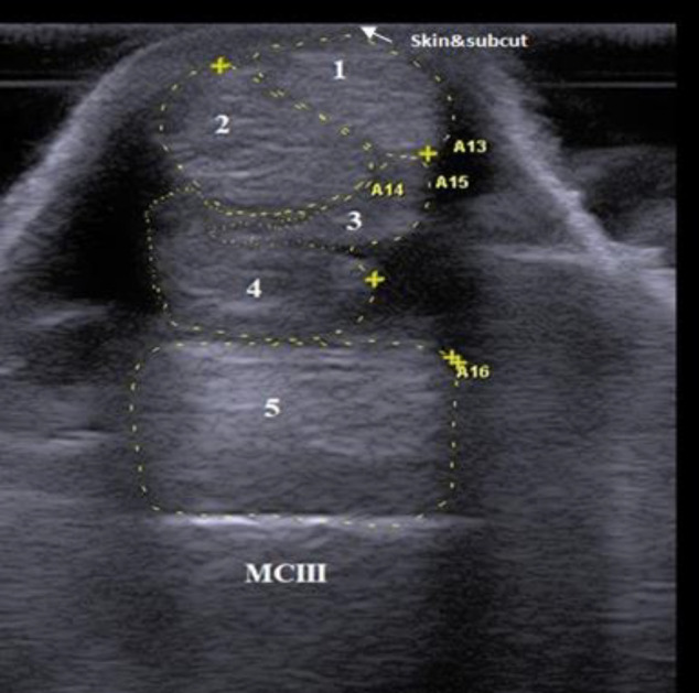
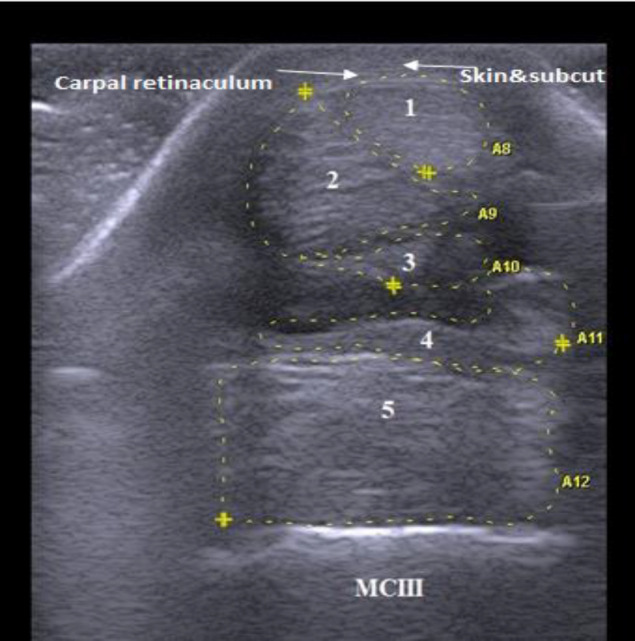
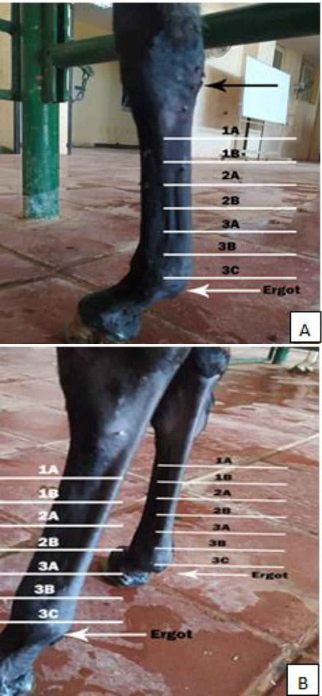
Methods: B-mode ultrasonography was conducted for the proposed tendons and ligaments in 12 clinically normal donkeys. Targeted structures were examined using transverse and longitudinal scans and evaluated in shape, echogenicity, echogenic pattern, fiber alignment pattern, and cross-sectional area (CSA).
Results: Using transverse scan, the sonographic shapes of tendons and ligaments of metacarpal and metatarsal regions were determined. Upon examining different levels of each region, specific ligaments, not tendons, were present only at the higher levels. The echogenicity of ligaments and tendons was either similar or variable across different levels. All tendons and ligaments displayed homogeneous echogenicity except for the suspensory ligament. In the longitudinal scan, tendons maintained linear and parallel fiber alignment along examination levels. Conversely, ligaments displayed mostly inconstant fiber patterns (linear/crimp). There was a statistically significant (P<0.05) difference in CSA of tendons and ligaments between certain levels within metacarpal and metatarsal regions. Upon comparing metacarpal and metatarsal regions, there were distinct variations in some ultrasonographic characteristics of the same tendons and ligaments.
Conclusion: This study established the ultrasonographic features of normal tendons and ligaments at the palmar/plantar aspect of the cannon region in donkeys (Equus asinus). These ultrasonographic data can be a reference guide when cannon region lameness is suspected.
期刊介绍:
The Iranian Journal of Veterinary Research(IJVR) is published quarterly in 4 issues. The aims of this journal are to improve and expand knowledge in all veterinary fields. It is an international journal indexed by the Thomson Institute for Scientific Information (ISI), Elsevier, Scopus, CAB International, Veterinary Bulletin and several other international databases. Research papers and reports on a wide range of veterinary topics are published in the journal after being evaluated by expert reviewers.The Editor-in-Chief is responsible for the editorial content of the journal—including peer-reviewed manuscripts—and the timing of its publication.
 求助内容:
求助内容: 应助结果提醒方式:


