Jonathan Bennett, George D Thornton, Christian Nitsche, Francisco F Gama, Nikoo Aziminia, Uzma Gul, Abhishek Shetye, Peter Kellman, Rhodri H Davies, James C Moon, Thomas A Treibel
{"title":"主动脉瓣置换术后2个月左心室肥厚:早期细胞和基质回归。","authors":"Jonathan Bennett, George D Thornton, Christian Nitsche, Francisco F Gama, Nikoo Aziminia, Uzma Gul, Abhishek Shetye, Peter Kellman, Rhodri H Davies, James C Moon, Thomas A Treibel","doi":"10.1161/CIRCIMAGING.124.017425","DOIUrl":null,"url":null,"abstract":"<p><strong>Background: </strong>In aortic stenosis, the myocardium responds with left ventricular hypertrophy, which is characterized by increased left ventricular mass due to cellular hypertrophy and extracellular matrix expansion. Following aortic valve replacement (AVR), left ventricular hypertrophy regression occurs, but early cellular and extracellular dynamics are unknown.</p><p><strong>Methods: </strong>Patients with severe symptomatic aortic stenosis undergoing surgical or transcatheter AVR were prospectively recruited. Pre- and early post-AVR cardiac magnetic resonance imaging assessed left ventricular remodeling, global longitudinal strain, and T1 mapping to determine extracellular volume fraction and volume of cellular and extracellular compartments.</p><p><strong>Results: </strong>In all, 39 patients (aged 71.4±9.8 years, male 79%, aortic valve peak velocity 4.4±0.5 m/s) underwent cardiac magnetic resonance before and at median 7.7 weeks post-AVR. Left ventricular mass index reduced significantly by 15.4% (<i>P</i><0.001*), primarily driven by cellular compartment regression (18.7%, <i>P</i><0.001*), with a smaller reduction in the extracellular compartment (7.2%, <i>P</i><0.001*). This unbalanced regression led to an apparent increase in extracellular volume fraction (27.4±3.1% to 30.2±2.8%; <i>P</i><0.001*). Although there was no significant change in global longitudinal strain post-AVR, an increase in extracellular volume fraction was associated with worsening of global longitudinal strain (Pearson r=0.41, <i>P</i>=0.01). Mode of intervention (transcatheter versus surgical) did not influence the above myocardial parameters post-AVR (all <i>P</i>>0.05). The asterisk in <i>P</i> values indicates a statistical significance of <0.05.</p><p><strong>Conclusions: </strong>Within 8 weeks of AVR for aortic stenosis, substantial left ventricular hypertrophy regression occurs involving both cellular and extracellular compartments, demonstrating the early myocardial adaptability to afterload relief. Cellular compartment regression is greater than extracellular regression, leading to an apparent increase in extracellular volume fraction. Mode of intervention did not affect degree of reverse remodeling, indicating that both are effective at resulting beneficial changes post-AVR.</p><p><strong>Registration: </strong>URL: https://www.isrctn.com; Unique identifier: NCT04627987.</p>","PeriodicalId":10202,"journal":{"name":"Circulation: Cardiovascular Imaging","volume":" ","pages":"e017425"},"PeriodicalIF":7.0000,"publicationDate":"2024-12-01","publicationTypes":"Journal Article","fieldsOfStudy":null,"isOpenAccess":false,"openAccessPdf":"https://www.ncbi.nlm.nih.gov/pmc/articles/PMC11649182/pdf/","citationCount":"0","resultStr":"{\"title\":\"Left Ventricular Hypertrophy in Aortic Stenosis: Early Cell and Matrix Regression 2 Months Post-Aortic Valve Replacement.\",\"authors\":\"Jonathan Bennett, George D Thornton, Christian Nitsche, Francisco F Gama, Nikoo Aziminia, Uzma Gul, Abhishek Shetye, Peter Kellman, Rhodri H Davies, James C Moon, Thomas A Treibel\",\"doi\":\"10.1161/CIRCIMAGING.124.017425\",\"DOIUrl\":null,\"url\":null,\"abstract\":\"<p><strong>Background: </strong>In aortic stenosis, the myocardium responds with left ventricular hypertrophy, which is characterized by increased left ventricular mass due to cellular hypertrophy and extracellular matrix expansion. Following aortic valve replacement (AVR), left ventricular hypertrophy regression occurs, but early cellular and extracellular dynamics are unknown.</p><p><strong>Methods: </strong>Patients with severe symptomatic aortic stenosis undergoing surgical or transcatheter AVR were prospectively recruited. Pre- and early post-AVR cardiac magnetic resonance imaging assessed left ventricular remodeling, global longitudinal strain, and T1 mapping to determine extracellular volume fraction and volume of cellular and extracellular compartments.</p><p><strong>Results: </strong>In all, 39 patients (aged 71.4±9.8 years, male 79%, aortic valve peak velocity 4.4±0.5 m/s) underwent cardiac magnetic resonance before and at median 7.7 weeks post-AVR. Left ventricular mass index reduced significantly by 15.4% (<i>P</i><0.001*), primarily driven by cellular compartment regression (18.7%, <i>P</i><0.001*), with a smaller reduction in the extracellular compartment (7.2%, <i>P</i><0.001*). This unbalanced regression led to an apparent increase in extracellular volume fraction (27.4±3.1% to 30.2±2.8%; <i>P</i><0.001*). Although there was no significant change in global longitudinal strain post-AVR, an increase in extracellular volume fraction was associated with worsening of global longitudinal strain (Pearson r=0.41, <i>P</i>=0.01). Mode of intervention (transcatheter versus surgical) did not influence the above myocardial parameters post-AVR (all <i>P</i>>0.05). The asterisk in <i>P</i> values indicates a statistical significance of <0.05.</p><p><strong>Conclusions: </strong>Within 8 weeks of AVR for aortic stenosis, substantial left ventricular hypertrophy regression occurs involving both cellular and extracellular compartments, demonstrating the early myocardial adaptability to afterload relief. Cellular compartment regression is greater than extracellular regression, leading to an apparent increase in extracellular volume fraction. Mode of intervention did not affect degree of reverse remodeling, indicating that both are effective at resulting beneficial changes post-AVR.</p><p><strong>Registration: </strong>URL: https://www.isrctn.com; Unique identifier: NCT04627987.</p>\",\"PeriodicalId\":10202,\"journal\":{\"name\":\"Circulation: Cardiovascular Imaging\",\"volume\":\" \",\"pages\":\"e017425\"},\"PeriodicalIF\":7.0000,\"publicationDate\":\"2024-12-01\",\"publicationTypes\":\"Journal Article\",\"fieldsOfStudy\":null,\"isOpenAccess\":false,\"openAccessPdf\":\"https://www.ncbi.nlm.nih.gov/pmc/articles/PMC11649182/pdf/\",\"citationCount\":\"0\",\"resultStr\":null,\"platform\":\"Semanticscholar\",\"paperid\":null,\"PeriodicalName\":\"Circulation: Cardiovascular Imaging\",\"FirstCategoryId\":\"3\",\"ListUrlMain\":\"https://doi.org/10.1161/CIRCIMAGING.124.017425\",\"RegionNum\":1,\"RegionCategory\":\"医学\",\"ArticlePicture\":[],\"TitleCN\":null,\"AbstractTextCN\":null,\"PMCID\":null,\"EPubDate\":\"2024/12/4 0:00:00\",\"PubModel\":\"Epub\",\"JCR\":\"Q1\",\"JCRName\":\"CARDIAC & CARDIOVASCULAR SYSTEMS\",\"Score\":null,\"Total\":0}","platform":"Semanticscholar","paperid":null,"PeriodicalName":"Circulation: Cardiovascular Imaging","FirstCategoryId":"3","ListUrlMain":"https://doi.org/10.1161/CIRCIMAGING.124.017425","RegionNum":1,"RegionCategory":"医学","ArticlePicture":[],"TitleCN":null,"AbstractTextCN":null,"PMCID":null,"EPubDate":"2024/12/4 0:00:00","PubModel":"Epub","JCR":"Q1","JCRName":"CARDIAC & CARDIOVASCULAR SYSTEMS","Score":null,"Total":0}
Left Ventricular Hypertrophy in Aortic Stenosis: Early Cell and Matrix Regression 2 Months Post-Aortic Valve Replacement.
Background: In aortic stenosis, the myocardium responds with left ventricular hypertrophy, which is characterized by increased left ventricular mass due to cellular hypertrophy and extracellular matrix expansion. Following aortic valve replacement (AVR), left ventricular hypertrophy regression occurs, but early cellular and extracellular dynamics are unknown.
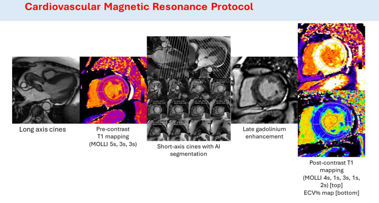
Methods: Patients with severe symptomatic aortic stenosis undergoing surgical or transcatheter AVR were prospectively recruited. Pre- and early post-AVR cardiac magnetic resonance imaging assessed left ventricular remodeling, global longitudinal strain, and T1 mapping to determine extracellular volume fraction and volume of cellular and extracellular compartments.
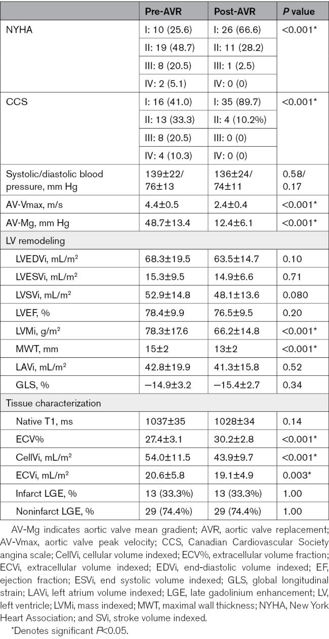
Results: In all, 39 patients (aged 71.4±9.8 years, male 79%, aortic valve peak velocity 4.4±0.5 m/s) underwent cardiac magnetic resonance before and at median 7.7 weeks post-AVR. Left ventricular mass index reduced significantly by 15.4% (P<0.001*), primarily driven by cellular compartment regression (18.7%, P<0.001*), with a smaller reduction in the extracellular compartment (7.2%, P<0.001*). This unbalanced regression led to an apparent increase in extracellular volume fraction (27.4±3.1% to 30.2±2.8%; P<0.001*). Although there was no significant change in global longitudinal strain post-AVR, an increase in extracellular volume fraction was associated with worsening of global longitudinal strain (Pearson r=0.41, P=0.01). Mode of intervention (transcatheter versus surgical) did not influence the above myocardial parameters post-AVR (all P>0.05). The asterisk in P values indicates a statistical significance of <0.05.
Conclusions: Within 8 weeks of AVR for aortic stenosis, substantial left ventricular hypertrophy regression occurs involving both cellular and extracellular compartments, demonstrating the early myocardial adaptability to afterload relief. Cellular compartment regression is greater than extracellular regression, leading to an apparent increase in extracellular volume fraction. Mode of intervention did not affect degree of reverse remodeling, indicating that both are effective at resulting beneficial changes post-AVR.
期刊介绍:
Circulation: Cardiovascular Imaging, an American Heart Association journal, publishes high-quality, patient-centric articles focusing on observational studies, clinical trials, and advances in applied (translational) research. The journal features innovative, multimodality approaches to the diagnosis and risk stratification of cardiovascular disease. Modalities covered include echocardiography, cardiac computed tomography, cardiac magnetic resonance imaging and spectroscopy, magnetic resonance angiography, cardiac positron emission tomography, noninvasive assessment of vascular and endothelial function, radionuclide imaging, molecular imaging, and others.
Article types considered by Circulation: Cardiovascular Imaging include Original Research, Research Letters, Advances in Cardiovascular Imaging, Clinical Implications of Molecular Imaging Research, How to Use Imaging, Translating Novel Imaging Technologies into Clinical Applications, and Cardiovascular Images.


 求助内容:
求助内容: 应助结果提醒方式:


