Colleen C Caldwell, Tinka V M Clement, Gijs J L Wuite
{"title":"人类有丝分裂中的力产生和阻力。","authors":"Colleen C Caldwell, Tinka V M Clement, Gijs J L Wuite","doi":"10.1007/s12551-024-01235-0","DOIUrl":null,"url":null,"abstract":"<p><p>Since the first observations of chromosome segregation over 150 years ago, efforts to observe the forces that drive mitosis have evolved alongside advances in microscopy. The mitotic spindle acts as the major generator of force through the highly regulated polymerization and depolymerization of microtubules as well as associated motor proteins. Centromeric chromatin, along with associated proteins including cohesin and condensin, is organized to resist these forces and ensure accurate chromosome segregation. Microtubules and centromeric chromatin join at the kinetochore, a complex protein superstructure. Ongoing research into the forces generated at the kinetochore-microtubule interface has resulted in a range of estimates for forces necessary to separate chromosomes, from tens to hundreds of piconewtons. Still, the exact magnitude and regulation of these forces remain areas of continuing investigation. Determining the precise forces involved in chromosome segregation is hindered by limitations of current measurement techniques, but advances such as optical tweezers combined with fluorescence microscopy are promising for future research.</p>","PeriodicalId":9094,"journal":{"name":"Biophysical reviews","volume":"16 5","pages":"551-562"},"PeriodicalIF":3.7000,"publicationDate":"2024-09-28","publicationTypes":"Journal Article","fieldsOfStudy":null,"isOpenAccess":false,"openAccessPdf":"https://www.ncbi.nlm.nih.gov/pmc/articles/PMC11604895/pdf/","citationCount":"0","resultStr":"{\"title\":\"Force generation and resistance in human mitosis.\",\"authors\":\"Colleen C Caldwell, Tinka V M Clement, Gijs J L Wuite\",\"doi\":\"10.1007/s12551-024-01235-0\",\"DOIUrl\":null,\"url\":null,\"abstract\":\"<p><p>Since the first observations of chromosome segregation over 150 years ago, efforts to observe the forces that drive mitosis have evolved alongside advances in microscopy. The mitotic spindle acts as the major generator of force through the highly regulated polymerization and depolymerization of microtubules as well as associated motor proteins. Centromeric chromatin, along with associated proteins including cohesin and condensin, is organized to resist these forces and ensure accurate chromosome segregation. Microtubules and centromeric chromatin join at the kinetochore, a complex protein superstructure. Ongoing research into the forces generated at the kinetochore-microtubule interface has resulted in a range of estimates for forces necessary to separate chromosomes, from tens to hundreds of piconewtons. Still, the exact magnitude and regulation of these forces remain areas of continuing investigation. Determining the precise forces involved in chromosome segregation is hindered by limitations of current measurement techniques, but advances such as optical tweezers combined with fluorescence microscopy are promising for future research.</p>\",\"PeriodicalId\":9094,\"journal\":{\"name\":\"Biophysical reviews\",\"volume\":\"16 5\",\"pages\":\"551-562\"},\"PeriodicalIF\":3.7000,\"publicationDate\":\"2024-09-28\",\"publicationTypes\":\"Journal Article\",\"fieldsOfStudy\":null,\"isOpenAccess\":false,\"openAccessPdf\":\"https://www.ncbi.nlm.nih.gov/pmc/articles/PMC11604895/pdf/\",\"citationCount\":\"0\",\"resultStr\":null,\"platform\":\"Semanticscholar\",\"paperid\":null,\"PeriodicalName\":\"Biophysical reviews\",\"FirstCategoryId\":\"1085\",\"ListUrlMain\":\"https://doi.org/10.1007/s12551-024-01235-0\",\"RegionNum\":0,\"RegionCategory\":null,\"ArticlePicture\":[],\"TitleCN\":null,\"AbstractTextCN\":null,\"PMCID\":null,\"EPubDate\":\"2024/10/1 0:00:00\",\"PubModel\":\"eCollection\",\"JCR\":\"Q1\",\"JCRName\":\"BIOPHYSICS\",\"Score\":null,\"Total\":0}","platform":"Semanticscholar","paperid":null,"PeriodicalName":"Biophysical reviews","FirstCategoryId":"1085","ListUrlMain":"https://doi.org/10.1007/s12551-024-01235-0","RegionNum":0,"RegionCategory":null,"ArticlePicture":[],"TitleCN":null,"AbstractTextCN":null,"PMCID":null,"EPubDate":"2024/10/1 0:00:00","PubModel":"eCollection","JCR":"Q1","JCRName":"BIOPHYSICS","Score":null,"Total":0}
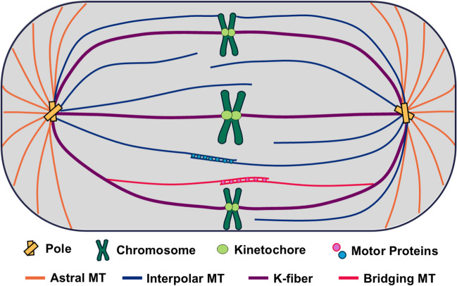
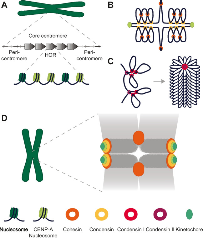
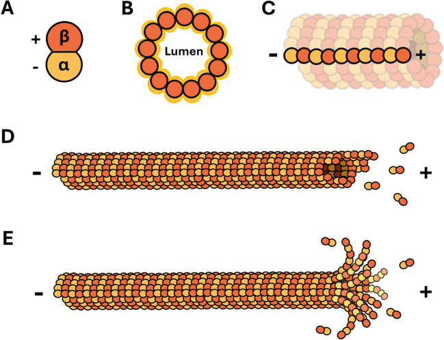
Since the first observations of chromosome segregation over 150 years ago, efforts to observe the forces that drive mitosis have evolved alongside advances in microscopy. The mitotic spindle acts as the major generator of force through the highly regulated polymerization and depolymerization of microtubules as well as associated motor proteins. Centromeric chromatin, along with associated proteins including cohesin and condensin, is organized to resist these forces and ensure accurate chromosome segregation. Microtubules and centromeric chromatin join at the kinetochore, a complex protein superstructure. Ongoing research into the forces generated at the kinetochore-microtubule interface has resulted in a range of estimates for forces necessary to separate chromosomes, from tens to hundreds of piconewtons. Still, the exact magnitude and regulation of these forces remain areas of continuing investigation. Determining the precise forces involved in chromosome segregation is hindered by limitations of current measurement techniques, but advances such as optical tweezers combined with fluorescence microscopy are promising for future research.
期刊介绍:
Biophysical Reviews aims to publish critical and timely reviews from key figures in the field of biophysics. The bulk of the reviews that are currently published are from invited authors, but the journal is also open for non-solicited reviews. Interested authors are encouraged to discuss the possibility of contributing a review with the Editor-in-Chief prior to submission. Through publishing reviews on biophysics, the editors of the journal hope to illustrate the great power and potential of physical techniques in the biological sciences, they aim to stimulate the discussion and promote further research and would like to educate and enthuse basic researcher scientists and students of biophysics. Biophysical Reviews covers the entire field of biophysics, generally defined as the science of describing and defining biological phenomenon using the concepts and the techniques of physics. This includes but is not limited by such areas as: - Bioinformatics - Biophysical methods and instrumentation - Medical biophysics - Biosystems - Cell biophysics and organization - Macromolecules: dynamics, structures and interactions - Single molecule biophysics - Membrane biophysics, channels and transportation

 求助内容:
求助内容: 应助结果提醒方式:


