Jichao Ma, Ariege Bizanti, Andrew M. Kwiat, Kayla Barton, Duyen Nguyen, Jazune Madas, Zulema Toledo, Kohlton Bendowski, Jin Chen, Zixi Jack Cheng
{"title":"大鼠全心房平丘左背根神经节的脊髓传入神经支配:顺行示踪","authors":"Jichao Ma, Ariege Bizanti, Andrew M. Kwiat, Kayla Barton, Duyen Nguyen, Jazune Madas, Zulema Toledo, Kohlton Bendowski, Jin Chen, Zixi Jack Cheng","doi":"10.1002/cne.25681","DOIUrl":null,"url":null,"abstract":"<div>\n \n <p>The spinal afferent innervation of the heart helps to regulate cardiac functions by sending sensory information through the dorsal root ganglia (DRG) to the brain. However, the distribution and morphology of spinal afferents in the heart are not well characterized due to tracer selections, the surgical access to upper thoracic DRGs, and the thickness of the heart tissues. In this study, we injected tracer dextran biotin (DB) into the left DRGs (C8-T3) of male Sprague–Dawley rats (3–5 months). After 16 days, flat-mounts of the whole left and right atria were prepared and diaminobenzidine stained. Then, the DB-labeled axons in the tissues were imaged, traced, and digitized using the Neurolucida system. Our results showed that the DB-labeled axons from left DRGs entered the left precaval vein and projected to the left and right atria, with predominant projection in the left atrial wall. DB-labeled varicose axons were observed in different layers, mostly in the epicardium and myocardium, but much less in the endocardium. In those layers, these spinal afferent axons branched out into simple to complex terminal arborizations, forming close appositions with cardiac muscles, intrinsic cardiac ganglia, blood vessels, and fat tissue. This work, for the first time, characterized cardiac spinal afferent distribution of the rat atria using anterograde tracing, which will provide the foundation for future studies of topographical cardiac spinal afferent innervation and remodeling in heart disease models.</p>\n </div>","PeriodicalId":15552,"journal":{"name":"Journal of Comparative Neurology","volume":"532 12","pages":""},"PeriodicalIF":2.1000,"publicationDate":"2024-12-02","publicationTypes":"Journal Article","fieldsOfStudy":null,"isOpenAccess":false,"openAccessPdf":"","citationCount":"0","resultStr":"{\"title\":\"Spinal Afferent Innervation From Left Dorsal Root Ganglia in the Flat-Mounts of Whole Atria of Rats: Anterograde Tracing\",\"authors\":\"Jichao Ma, Ariege Bizanti, Andrew M. Kwiat, Kayla Barton, Duyen Nguyen, Jazune Madas, Zulema Toledo, Kohlton Bendowski, Jin Chen, Zixi Jack Cheng\",\"doi\":\"10.1002/cne.25681\",\"DOIUrl\":null,\"url\":null,\"abstract\":\"<div>\\n \\n <p>The spinal afferent innervation of the heart helps to regulate cardiac functions by sending sensory information through the dorsal root ganglia (DRG) to the brain. However, the distribution and morphology of spinal afferents in the heart are not well characterized due to tracer selections, the surgical access to upper thoracic DRGs, and the thickness of the heart tissues. In this study, we injected tracer dextran biotin (DB) into the left DRGs (C8-T3) of male Sprague–Dawley rats (3–5 months). After 16 days, flat-mounts of the whole left and right atria were prepared and diaminobenzidine stained. Then, the DB-labeled axons in the tissues were imaged, traced, and digitized using the Neurolucida system. Our results showed that the DB-labeled axons from left DRGs entered the left precaval vein and projected to the left and right atria, with predominant projection in the left atrial wall. DB-labeled varicose axons were observed in different layers, mostly in the epicardium and myocardium, but much less in the endocardium. In those layers, these spinal afferent axons branched out into simple to complex terminal arborizations, forming close appositions with cardiac muscles, intrinsic cardiac ganglia, blood vessels, and fat tissue. This work, for the first time, characterized cardiac spinal afferent distribution of the rat atria using anterograde tracing, which will provide the foundation for future studies of topographical cardiac spinal afferent innervation and remodeling in heart disease models.</p>\\n </div>\",\"PeriodicalId\":15552,\"journal\":{\"name\":\"Journal of Comparative Neurology\",\"volume\":\"532 12\",\"pages\":\"\"},\"PeriodicalIF\":2.1000,\"publicationDate\":\"2024-12-02\",\"publicationTypes\":\"Journal Article\",\"fieldsOfStudy\":null,\"isOpenAccess\":false,\"openAccessPdf\":\"\",\"citationCount\":\"0\",\"resultStr\":null,\"platform\":\"Semanticscholar\",\"paperid\":null,\"PeriodicalName\":\"Journal of Comparative Neurology\",\"FirstCategoryId\":\"3\",\"ListUrlMain\":\"https://onlinelibrary.wiley.com/doi/10.1002/cne.25681\",\"RegionNum\":4,\"RegionCategory\":\"医学\",\"ArticlePicture\":[],\"TitleCN\":null,\"AbstractTextCN\":null,\"PMCID\":null,\"EPubDate\":\"\",\"PubModel\":\"\",\"JCR\":\"Q3\",\"JCRName\":\"NEUROSCIENCES\",\"Score\":null,\"Total\":0}","platform":"Semanticscholar","paperid":null,"PeriodicalName":"Journal of Comparative Neurology","FirstCategoryId":"3","ListUrlMain":"https://onlinelibrary.wiley.com/doi/10.1002/cne.25681","RegionNum":4,"RegionCategory":"医学","ArticlePicture":[],"TitleCN":null,"AbstractTextCN":null,"PMCID":null,"EPubDate":"","PubModel":"","JCR":"Q3","JCRName":"NEUROSCIENCES","Score":null,"Total":0}
Spinal Afferent Innervation From Left Dorsal Root Ganglia in the Flat-Mounts of Whole Atria of Rats: Anterograde Tracing
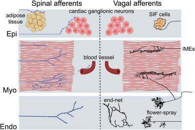
The spinal afferent innervation of the heart helps to regulate cardiac functions by sending sensory information through the dorsal root ganglia (DRG) to the brain. However, the distribution and morphology of spinal afferents in the heart are not well characterized due to tracer selections, the surgical access to upper thoracic DRGs, and the thickness of the heart tissues. In this study, we injected tracer dextran biotin (DB) into the left DRGs (C8-T3) of male Sprague–Dawley rats (3–5 months). After 16 days, flat-mounts of the whole left and right atria were prepared and diaminobenzidine stained. Then, the DB-labeled axons in the tissues were imaged, traced, and digitized using the Neurolucida system. Our results showed that the DB-labeled axons from left DRGs entered the left precaval vein and projected to the left and right atria, with predominant projection in the left atrial wall. DB-labeled varicose axons were observed in different layers, mostly in the epicardium and myocardium, but much less in the endocardium. In those layers, these spinal afferent axons branched out into simple to complex terminal arborizations, forming close appositions with cardiac muscles, intrinsic cardiac ganglia, blood vessels, and fat tissue. This work, for the first time, characterized cardiac spinal afferent distribution of the rat atria using anterograde tracing, which will provide the foundation for future studies of topographical cardiac spinal afferent innervation and remodeling in heart disease models.
期刊介绍:
Established in 1891, JCN is the oldest continually published basic neuroscience journal. Historically, as the name suggests, the journal focused on a comparison among species to uncover the intricacies of how the brain functions. In modern times, this research is called systems neuroscience where animal models are used to mimic core cognitive processes with the ultimate goal of understanding neural circuits and connections that give rise to behavioral patterns and different neural states.
Research published in JCN covers all species from invertebrates to humans, and the reports inform the readers about the function and organization of nervous systems in species with an emphasis on the way that species adaptations inform about the function or organization of the nervous systems, rather than on their evolution per se.
JCN publishes primary research articles and critical commentaries and review-type articles offering expert insight in to cutting edge research in the field of systems neuroscience; a complete list of contribution types is given in the Author Guidelines. For primary research contributions, only full-length investigative reports are desired; the journal does not accept short communications.

 求助内容:
求助内容: 应助结果提醒方式:


