Michael Tseng, Avrey Thau, Carla Berkowitz, Abhijit Ramaprasad, Surendra Basti
{"title":"术中光学相干断层成像评估前房气体充盈。","authors":"Michael Tseng, Avrey Thau, Carla Berkowitz, Abhijit Ramaprasad, Surendra Basti","doi":"10.3389/fopht.2024.1488764","DOIUrl":null,"url":null,"abstract":"<p><strong>Introduction: </strong>During endothelial keratoplasty, anterior chamber gas is titrated to a desired fill, which is difficult to optimize by visualization alone. This study evaluates how an anterior chamber gas fill correlates with intraocular pressure (IOP) and iris-angle configuration as identified by optical coherence tomography (OCT).</p><p><strong>Methods: </strong>Eleven cadaveric eyes were studied in three configurations: baseline, air-fill just spanning limbus-to-limbus (\"full-fill\"), and air-fill maximally filling the anterior chamber (\"overfill\"). At each configuration, IOP was measured by Tonopen and iris-angle was determined by analyzing OCT images.</p><p><strong>Results: </strong>No differences in IOP or irisangles were identified between baseline and full-fill configurations (p=0.113 and p=0.152, respectively). When compared to overfill configuration, differences in IOP and iris-angles were identified for baseline (p<0.001 and p=0.001, respectively) and full-fill configuration (p=0.001 and p=0.039, respectively).</p><p><strong>Discussion: </strong>These findings highlight that en-face visualization of full-fill may not be indicative of IOP elevation. A significant difference in IOP and iris-angle exists between full-fill and overfill configurations. Intraoperative OCT can serve as a useful surrogate to identify the extent of fill.</p>","PeriodicalId":73096,"journal":{"name":"Frontiers in ophthalmology","volume":"4 ","pages":"1488764"},"PeriodicalIF":0.9000,"publicationDate":"2024-11-14","publicationTypes":"Journal Article","fieldsOfStudy":null,"isOpenAccess":false,"openAccessPdf":"https://www.ncbi.nlm.nih.gov/pmc/articles/PMC11602447/pdf/","citationCount":"0","resultStr":"{\"title\":\"Intraoperative optical coherence tomography imaging for assessment of anterior chamber gas fill.\",\"authors\":\"Michael Tseng, Avrey Thau, Carla Berkowitz, Abhijit Ramaprasad, Surendra Basti\",\"doi\":\"10.3389/fopht.2024.1488764\",\"DOIUrl\":null,\"url\":null,\"abstract\":\"<p><strong>Introduction: </strong>During endothelial keratoplasty, anterior chamber gas is titrated to a desired fill, which is difficult to optimize by visualization alone. This study evaluates how an anterior chamber gas fill correlates with intraocular pressure (IOP) and iris-angle configuration as identified by optical coherence tomography (OCT).</p><p><strong>Methods: </strong>Eleven cadaveric eyes were studied in three configurations: baseline, air-fill just spanning limbus-to-limbus (\\\"full-fill\\\"), and air-fill maximally filling the anterior chamber (\\\"overfill\\\"). At each configuration, IOP was measured by Tonopen and iris-angle was determined by analyzing OCT images.</p><p><strong>Results: </strong>No differences in IOP or irisangles were identified between baseline and full-fill configurations (p=0.113 and p=0.152, respectively). When compared to overfill configuration, differences in IOP and iris-angles were identified for baseline (p<0.001 and p=0.001, respectively) and full-fill configuration (p=0.001 and p=0.039, respectively).</p><p><strong>Discussion: </strong>These findings highlight that en-face visualization of full-fill may not be indicative of IOP elevation. A significant difference in IOP and iris-angle exists between full-fill and overfill configurations. Intraoperative OCT can serve as a useful surrogate to identify the extent of fill.</p>\",\"PeriodicalId\":73096,\"journal\":{\"name\":\"Frontiers in ophthalmology\",\"volume\":\"4 \",\"pages\":\"1488764\"},\"PeriodicalIF\":0.9000,\"publicationDate\":\"2024-11-14\",\"publicationTypes\":\"Journal Article\",\"fieldsOfStudy\":null,\"isOpenAccess\":false,\"openAccessPdf\":\"https://www.ncbi.nlm.nih.gov/pmc/articles/PMC11602447/pdf/\",\"citationCount\":\"0\",\"resultStr\":null,\"platform\":\"Semanticscholar\",\"paperid\":null,\"PeriodicalName\":\"Frontiers in ophthalmology\",\"FirstCategoryId\":\"1085\",\"ListUrlMain\":\"https://doi.org/10.3389/fopht.2024.1488764\",\"RegionNum\":0,\"RegionCategory\":null,\"ArticlePicture\":[],\"TitleCN\":null,\"AbstractTextCN\":null,\"PMCID\":null,\"EPubDate\":\"2024/1/1 0:00:00\",\"PubModel\":\"eCollection\",\"JCR\":\"\",\"JCRName\":\"\",\"Score\":null,\"Total\":0}","platform":"Semanticscholar","paperid":null,"PeriodicalName":"Frontiers in ophthalmology","FirstCategoryId":"1085","ListUrlMain":"https://doi.org/10.3389/fopht.2024.1488764","RegionNum":0,"RegionCategory":null,"ArticlePicture":[],"TitleCN":null,"AbstractTextCN":null,"PMCID":null,"EPubDate":"2024/1/1 0:00:00","PubModel":"eCollection","JCR":"","JCRName":"","Score":null,"Total":0}
Intraoperative optical coherence tomography imaging for assessment of anterior chamber gas fill.
Introduction: During endothelial keratoplasty, anterior chamber gas is titrated to a desired fill, which is difficult to optimize by visualization alone. This study evaluates how an anterior chamber gas fill correlates with intraocular pressure (IOP) and iris-angle configuration as identified by optical coherence tomography (OCT).
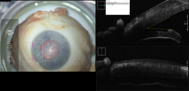
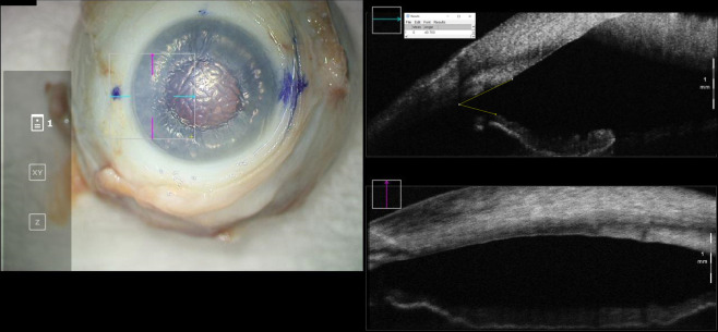
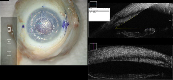
Methods: Eleven cadaveric eyes were studied in three configurations: baseline, air-fill just spanning limbus-to-limbus ("full-fill"), and air-fill maximally filling the anterior chamber ("overfill"). At each configuration, IOP was measured by Tonopen and iris-angle was determined by analyzing OCT images.
Results: No differences in IOP or irisangles were identified between baseline and full-fill configurations (p=0.113 and p=0.152, respectively). When compared to overfill configuration, differences in IOP and iris-angles were identified for baseline (p<0.001 and p=0.001, respectively) and full-fill configuration (p=0.001 and p=0.039, respectively).
Discussion: These findings highlight that en-face visualization of full-fill may not be indicative of IOP elevation. A significant difference in IOP and iris-angle exists between full-fill and overfill configurations. Intraoperative OCT can serve as a useful surrogate to identify the extent of fill.

 求助内容:
求助内容: 应助结果提醒方式:


