Ephraim A. Ansa-Addo, Paras Pathak, Maria V. McCrossan, Izadora Volpato Rossi, Mahamed Abdullahi, Dan Stratton, Sigrun Lange, Marcel I. Ramirez, Jameel M. Inal
{"title":"克氏锥虫刺激单核细胞来源的细胞外囊泡,通过激活TGF-β1增强细胞体外侵袭","authors":"Ephraim A. Ansa-Addo, Paras Pathak, Maria V. McCrossan, Izadora Volpato Rossi, Mahamed Abdullahi, Dan Stratton, Sigrun Lange, Marcel I. Ramirez, Jameel M. Inal","doi":"10.1002/jev2.70014","DOIUrl":null,"url":null,"abstract":"<p>During cell invasion, large Extracellular Vesicle (lEV) release from host cells was dose-dependently triggered by <i>Trypanosoma cruzi</i> metacyclic trypomastigotes (Mtr). This lEV release was inhibited when IP<sub>3</sub>-mediated Ca<sup>2+</sup> exit from the ER and further Ca<sup>2+</sup> entry from plasma membrane channels was blocked, but whilst any store-independent Ca<sup>2+</sup> entry (SICE) could continue unabated. That lEV release was equally inhibited if all entry from external sources was blocked by chelation of external Ca<sup>2+</sup> points to the major contributor to Mtr-triggered host cell lEV release being IP<sub>3</sub>/store-mediated Ca<sup>2+</sup> release, SICE playing a minor role. Host cell lEVs were released through Mtr interaction with host cell lipid raft domains, integrins, and mechanosensitive ion channels, whereupon [Ca<sup>2+</sup>]<sub>cyt</sub> increased (50 to 750 nM) within 15 s. lEV release and cell entry of <i>T. cruzi</i>, which increased up to 30 and 60 mpi, respectively, as well as raised actin depolymerization at 60 mpi, were all reduced by TRPC inhibitor, GsMTx-4. Vesicle release and infection was also reduced with RGD peptide, methyl-β-cyclodextrin, knockdown of calpain and with the calpain inhibitor, calpeptin. Restoration of lEV levels, whether with lEVs from infected or uninfected epithelial cells, did not restore invasion, but supplementation with lEVs from infected monocytes, did. We provide evidence of THP-1 monocyte-derived lEV interaction with Mtr (lipid mixing by R18-dequenching; flow cytometry showing transfer to Mtr of R18 from R18-lEVs and of LAP(TGF-β1). Active, mature TGF-β1 (at 175 pg/×10<sup>5</sup> in THP-1 lEVs) was detected in concentrated lEV-/cell-free supernatant by western blotting, only after THP-1 lEVs had interacted with Mtr. The TGF-β1 receptor (TβRI) inhibitor, SB-431542, reduced the enhanced cellular invasion due to monocyte-lEVs.</p>","PeriodicalId":15811,"journal":{"name":"Journal of Extracellular Vesicles","volume":"13 11","pages":""},"PeriodicalIF":14.5000,"publicationDate":"2024-11-29","publicationTypes":"Journal Article","fieldsOfStudy":null,"isOpenAccess":false,"openAccessPdf":"https://onlinelibrary.wiley.com/doi/epdf/10.1002/jev2.70014","citationCount":"0","resultStr":"{\"title\":\"Monocyte-derived extracellular vesicles, stimulated by Trypanosoma cruzi, enhance cellular invasion in vitro via activated TGF-β1\",\"authors\":\"Ephraim A. Ansa-Addo, Paras Pathak, Maria V. McCrossan, Izadora Volpato Rossi, Mahamed Abdullahi, Dan Stratton, Sigrun Lange, Marcel I. Ramirez, Jameel M. Inal\",\"doi\":\"10.1002/jev2.70014\",\"DOIUrl\":null,\"url\":null,\"abstract\":\"<p>During cell invasion, large Extracellular Vesicle (lEV) release from host cells was dose-dependently triggered by <i>Trypanosoma cruzi</i> metacyclic trypomastigotes (Mtr). This lEV release was inhibited when IP<sub>3</sub>-mediated Ca<sup>2+</sup> exit from the ER and further Ca<sup>2+</sup> entry from plasma membrane channels was blocked, but whilst any store-independent Ca<sup>2+</sup> entry (SICE) could continue unabated. That lEV release was equally inhibited if all entry from external sources was blocked by chelation of external Ca<sup>2+</sup> points to the major contributor to Mtr-triggered host cell lEV release being IP<sub>3</sub>/store-mediated Ca<sup>2+</sup> release, SICE playing a minor role. Host cell lEVs were released through Mtr interaction with host cell lipid raft domains, integrins, and mechanosensitive ion channels, whereupon [Ca<sup>2+</sup>]<sub>cyt</sub> increased (50 to 750 nM) within 15 s. lEV release and cell entry of <i>T. cruzi</i>, which increased up to 30 and 60 mpi, respectively, as well as raised actin depolymerization at 60 mpi, were all reduced by TRPC inhibitor, GsMTx-4. Vesicle release and infection was also reduced with RGD peptide, methyl-β-cyclodextrin, knockdown of calpain and with the calpain inhibitor, calpeptin. Restoration of lEV levels, whether with lEVs from infected or uninfected epithelial cells, did not restore invasion, but supplementation with lEVs from infected monocytes, did. We provide evidence of THP-1 monocyte-derived lEV interaction with Mtr (lipid mixing by R18-dequenching; flow cytometry showing transfer to Mtr of R18 from R18-lEVs and of LAP(TGF-β1). Active, mature TGF-β1 (at 175 pg/×10<sup>5</sup> in THP-1 lEVs) was detected in concentrated lEV-/cell-free supernatant by western blotting, only after THP-1 lEVs had interacted with Mtr. The TGF-β1 receptor (TβRI) inhibitor, SB-431542, reduced the enhanced cellular invasion due to monocyte-lEVs.</p>\",\"PeriodicalId\":15811,\"journal\":{\"name\":\"Journal of Extracellular Vesicles\",\"volume\":\"13 11\",\"pages\":\"\"},\"PeriodicalIF\":14.5000,\"publicationDate\":\"2024-11-29\",\"publicationTypes\":\"Journal Article\",\"fieldsOfStudy\":null,\"isOpenAccess\":false,\"openAccessPdf\":\"https://onlinelibrary.wiley.com/doi/epdf/10.1002/jev2.70014\",\"citationCount\":\"0\",\"resultStr\":null,\"platform\":\"Semanticscholar\",\"paperid\":null,\"PeriodicalName\":\"Journal of Extracellular Vesicles\",\"FirstCategoryId\":\"3\",\"ListUrlMain\":\"https://onlinelibrary.wiley.com/doi/10.1002/jev2.70014\",\"RegionNum\":1,\"RegionCategory\":\"医学\",\"ArticlePicture\":[],\"TitleCN\":null,\"AbstractTextCN\":null,\"PMCID\":null,\"EPubDate\":\"\",\"PubModel\":\"\",\"JCR\":\"Q1\",\"JCRName\":\"CELL BIOLOGY\",\"Score\":null,\"Total\":0}","platform":"Semanticscholar","paperid":null,"PeriodicalName":"Journal of Extracellular Vesicles","FirstCategoryId":"3","ListUrlMain":"https://onlinelibrary.wiley.com/doi/10.1002/jev2.70014","RegionNum":1,"RegionCategory":"医学","ArticlePicture":[],"TitleCN":null,"AbstractTextCN":null,"PMCID":null,"EPubDate":"","PubModel":"","JCR":"Q1","JCRName":"CELL BIOLOGY","Score":null,"Total":0}
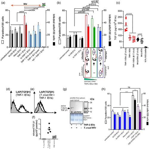
Monocyte-derived extracellular vesicles, stimulated by Trypanosoma cruzi, enhance cellular invasion in vitro via activated TGF-β1
During cell invasion, large Extracellular Vesicle (lEV) release from host cells was dose-dependently triggered by Trypanosoma cruzi metacyclic trypomastigotes (Mtr). This lEV release was inhibited when IP3-mediated Ca2+ exit from the ER and further Ca2+ entry from plasma membrane channels was blocked, but whilst any store-independent Ca2+ entry (SICE) could continue unabated. That lEV release was equally inhibited if all entry from external sources was blocked by chelation of external Ca2+ points to the major contributor to Mtr-triggered host cell lEV release being IP3/store-mediated Ca2+ release, SICE playing a minor role. Host cell lEVs were released through Mtr interaction with host cell lipid raft domains, integrins, and mechanosensitive ion channels, whereupon [Ca2+]cyt increased (50 to 750 nM) within 15 s. lEV release and cell entry of T. cruzi, which increased up to 30 and 60 mpi, respectively, as well as raised actin depolymerization at 60 mpi, were all reduced by TRPC inhibitor, GsMTx-4. Vesicle release and infection was also reduced with RGD peptide, methyl-β-cyclodextrin, knockdown of calpain and with the calpain inhibitor, calpeptin. Restoration of lEV levels, whether with lEVs from infected or uninfected epithelial cells, did not restore invasion, but supplementation with lEVs from infected monocytes, did. We provide evidence of THP-1 monocyte-derived lEV interaction with Mtr (lipid mixing by R18-dequenching; flow cytometry showing transfer to Mtr of R18 from R18-lEVs and of LAP(TGF-β1). Active, mature TGF-β1 (at 175 pg/×105 in THP-1 lEVs) was detected in concentrated lEV-/cell-free supernatant by western blotting, only after THP-1 lEVs had interacted with Mtr. The TGF-β1 receptor (TβRI) inhibitor, SB-431542, reduced the enhanced cellular invasion due to monocyte-lEVs.
期刊介绍:
The Journal of Extracellular Vesicles is an open access research publication that focuses on extracellular vesicles, including microvesicles, exosomes, ectosomes, and apoptotic bodies. It serves as the official journal of the International Society for Extracellular Vesicles and aims to facilitate the exchange of data, ideas, and information pertaining to the chemistry, biology, and applications of extracellular vesicles. The journal covers various aspects such as the cellular and molecular mechanisms of extracellular vesicles biogenesis, technological advancements in their isolation, quantification, and characterization, the role and function of extracellular vesicles in biology, stem cell-derived extracellular vesicles and their biology, as well as the application of extracellular vesicles for pharmacological, immunological, or genetic therapies.
The Journal of Extracellular Vesicles is widely recognized and indexed by numerous services, including Biological Abstracts, BIOSIS Previews, Chemical Abstracts Service (CAS), Current Contents/Life Sciences, Directory of Open Access Journals (DOAJ), Journal Citation Reports/Science Edition, Google Scholar, ProQuest Natural Science Collection, ProQuest SciTech Collection, SciTech Premium Collection, PubMed Central/PubMed, Science Citation Index Expanded, ScienceOpen, and Scopus.

 求助内容:
求助内容: 应助结果提醒方式:


