{"title":"胰岛细胞中多泡体和分泌颗粒的定位和释放的比较:2型糖尿病的失调","authors":"Priyadarshini Veerabhadraswamy, Kiran Lata, Sristi Dey, Prajakta Belekar, Lakshmi Kothegala, Vidya Mangala Prasad, Nikhil R. Gandasi","doi":"10.1002/jex2.70014","DOIUrl":null,"url":null,"abstract":"<p>Multivesicular bodies (MVBs) are vesicles of endosomal origin containing intraluminal vesicles, which upon fusion with plasma membrane, secrete exosomes. They play a significant role in the physiology and pathology of type-2 diabetes (T2D) due to disrupted intercellular communication. The role of MVBs and their influence on insulin secretory granules (ISGs) of β-cells or their characterization is yet to be uncovered. In our study, we compared MVBs to largely well-characterized ISGs in β-cells. This study compares the density, localization, and exocytosis of CD63+ compartments (CD63+c) with NPY labelled ISGs (NISGs) in β-cells. For this, tetraspanin CD63 was exploited to majorly label MVBs in β-cells. These labels preserve the structural integrity of labelled compartments and mostly do not localize with other endo-lysosomal compartments. This study showed that the β-cells have a significantly higher density of NISGs than CD63+c. CD63+c and NISGs are spatially localized apart within β-cells. The proteins that localize with CD63+c are different from the ones that localize with NISGs. Exocytosis of NISGs occurs at the periphery of the β-cells and takes significantly less time when compared to the release of CD63+c, which is non-peripheral and takes a longer duration. Mechanistically, the availability of CD63+c for exocytosis was assessed and found that an equilibrium is maintained between docking and undocking states at the plasma membrane. Although there are a high number of short-term residing, visiting CD63+c at the plasma membrane, the availability of CD63+c for exocytosis is maintained due to docking and undocking states. Further, a significant reduction in the density of NISGs and CD63+c was observed in β-cells isolated from T2D donors compared to healthy counterparts. Studying the effect of MVBs on insulin secretion in physiological and T2D conditions has huge potential. This study provides a strong basis to open new avenues for such future studies.</p>","PeriodicalId":73747,"journal":{"name":"Journal of extracellular biology","volume":"3 11","pages":""},"PeriodicalIF":0.0000,"publicationDate":"2024-11-29","publicationTypes":"Journal Article","fieldsOfStudy":null,"isOpenAccess":false,"openAccessPdf":"https://onlinelibrary.wiley.com/doi/epdf/10.1002/jex2.70014","citationCount":"0","resultStr":"{\"title\":\"Comparison of localization and release of multivesicular bodies and secretory granules in islet cells: Dysregulation during type-2 diabetes\",\"authors\":\"Priyadarshini Veerabhadraswamy, Kiran Lata, Sristi Dey, Prajakta Belekar, Lakshmi Kothegala, Vidya Mangala Prasad, Nikhil R. Gandasi\",\"doi\":\"10.1002/jex2.70014\",\"DOIUrl\":null,\"url\":null,\"abstract\":\"<p>Multivesicular bodies (MVBs) are vesicles of endosomal origin containing intraluminal vesicles, which upon fusion with plasma membrane, secrete exosomes. They play a significant role in the physiology and pathology of type-2 diabetes (T2D) due to disrupted intercellular communication. The role of MVBs and their influence on insulin secretory granules (ISGs) of β-cells or their characterization is yet to be uncovered. In our study, we compared MVBs to largely well-characterized ISGs in β-cells. This study compares the density, localization, and exocytosis of CD63+ compartments (CD63+c) with NPY labelled ISGs (NISGs) in β-cells. For this, tetraspanin CD63 was exploited to majorly label MVBs in β-cells. These labels preserve the structural integrity of labelled compartments and mostly do not localize with other endo-lysosomal compartments. This study showed that the β-cells have a significantly higher density of NISGs than CD63+c. CD63+c and NISGs are spatially localized apart within β-cells. The proteins that localize with CD63+c are different from the ones that localize with NISGs. Exocytosis of NISGs occurs at the periphery of the β-cells and takes significantly less time when compared to the release of CD63+c, which is non-peripheral and takes a longer duration. Mechanistically, the availability of CD63+c for exocytosis was assessed and found that an equilibrium is maintained between docking and undocking states at the plasma membrane. Although there are a high number of short-term residing, visiting CD63+c at the plasma membrane, the availability of CD63+c for exocytosis is maintained due to docking and undocking states. Further, a significant reduction in the density of NISGs and CD63+c was observed in β-cells isolated from T2D donors compared to healthy counterparts. Studying the effect of MVBs on insulin secretion in physiological and T2D conditions has huge potential. This study provides a strong basis to open new avenues for such future studies.</p>\",\"PeriodicalId\":73747,\"journal\":{\"name\":\"Journal of extracellular biology\",\"volume\":\"3 11\",\"pages\":\"\"},\"PeriodicalIF\":0.0000,\"publicationDate\":\"2024-11-29\",\"publicationTypes\":\"Journal Article\",\"fieldsOfStudy\":null,\"isOpenAccess\":false,\"openAccessPdf\":\"https://onlinelibrary.wiley.com/doi/epdf/10.1002/jex2.70014\",\"citationCount\":\"0\",\"resultStr\":null,\"platform\":\"Semanticscholar\",\"paperid\":null,\"PeriodicalName\":\"Journal of extracellular biology\",\"FirstCategoryId\":\"1085\",\"ListUrlMain\":\"https://isevjournals.onlinelibrary.wiley.com/doi/10.1002/jex2.70014\",\"RegionNum\":0,\"RegionCategory\":null,\"ArticlePicture\":[],\"TitleCN\":null,\"AbstractTextCN\":null,\"PMCID\":null,\"EPubDate\":\"\",\"PubModel\":\"\",\"JCR\":\"\",\"JCRName\":\"\",\"Score\":null,\"Total\":0}","platform":"Semanticscholar","paperid":null,"PeriodicalName":"Journal of extracellular biology","FirstCategoryId":"1085","ListUrlMain":"https://isevjournals.onlinelibrary.wiley.com/doi/10.1002/jex2.70014","RegionNum":0,"RegionCategory":null,"ArticlePicture":[],"TitleCN":null,"AbstractTextCN":null,"PMCID":null,"EPubDate":"","PubModel":"","JCR":"","JCRName":"","Score":null,"Total":0}
Comparison of localization and release of multivesicular bodies and secretory granules in islet cells: Dysregulation during type-2 diabetes
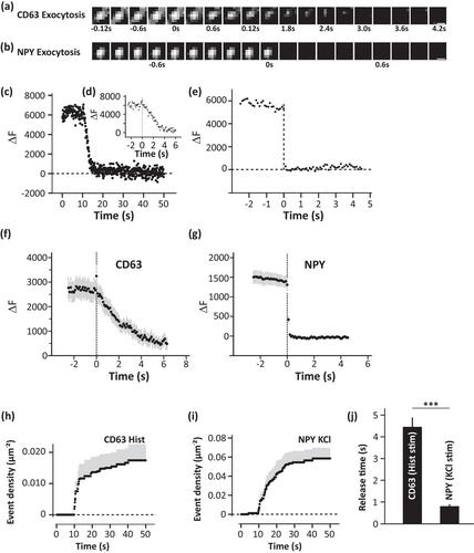
Multivesicular bodies (MVBs) are vesicles of endosomal origin containing intraluminal vesicles, which upon fusion with plasma membrane, secrete exosomes. They play a significant role in the physiology and pathology of type-2 diabetes (T2D) due to disrupted intercellular communication. The role of MVBs and their influence on insulin secretory granules (ISGs) of β-cells or their characterization is yet to be uncovered. In our study, we compared MVBs to largely well-characterized ISGs in β-cells. This study compares the density, localization, and exocytosis of CD63+ compartments (CD63+c) with NPY labelled ISGs (NISGs) in β-cells. For this, tetraspanin CD63 was exploited to majorly label MVBs in β-cells. These labels preserve the structural integrity of labelled compartments and mostly do not localize with other endo-lysosomal compartments. This study showed that the β-cells have a significantly higher density of NISGs than CD63+c. CD63+c and NISGs are spatially localized apart within β-cells. The proteins that localize with CD63+c are different from the ones that localize with NISGs. Exocytosis of NISGs occurs at the periphery of the β-cells and takes significantly less time when compared to the release of CD63+c, which is non-peripheral and takes a longer duration. Mechanistically, the availability of CD63+c for exocytosis was assessed and found that an equilibrium is maintained between docking and undocking states at the plasma membrane. Although there are a high number of short-term residing, visiting CD63+c at the plasma membrane, the availability of CD63+c for exocytosis is maintained due to docking and undocking states. Further, a significant reduction in the density of NISGs and CD63+c was observed in β-cells isolated from T2D donors compared to healthy counterparts. Studying the effect of MVBs on insulin secretion in physiological and T2D conditions has huge potential. This study provides a strong basis to open new avenues for such future studies.


 求助内容:
求助内容: 应助结果提醒方式:


