{"title":"正常解剖及内耳畸形的耳蜗神经显像","authors":"Majed Assiri MD, Tawfiq Khurayzi MD, Fida Almuhawas MD, Kurt Schlemmer MD, Abdulrahman Hagr MD, Anandhan Dhanasingh PhD","doi":"10.1002/lio2.70023","DOIUrl":null,"url":null,"abstract":"<div>\n \n \n <section>\n \n <h3> Objectives</h3>\n \n <p>This study aimed to qualitatively evaluate the variations in nerve bundles between patients with normal anatomy and those with inner-ear anomalies.</p>\n </section>\n \n <section>\n \n <h3> Methods</h3>\n \n <p>Magnetic resonance imaging (MRI) scans of the temporal bones of candidates for cochlear implants (CIs) enrolled at a tertiary center were retrospectively reviewed from the clinical database. The 3.0-Tesla MRI scans were analyzed using a three-dimensional slicer to visualize the nerve bundles in the internal auditory canal.</p>\n </section>\n \n <section>\n \n <h3> Results</h3>\n \n <p>A total of 49 ears were analyzed. Twenty ears exhibited normal inner ear anatomy, whereas 29 ears had various inner-ear malformations. The cochlear nerve (CN) was visible on all 20 scans with normal inner-ear anatomy. In addition, the CN was visualized in 18 scans with inner ear malformations. Furthermore, the CN was identified in six of the eight scans with IP type I, whereas in two scans, the CN and vestibular nerve were unclear. Three scans with a common cavity showed only two nerve bundles.</p>\n </section>\n \n <section>\n \n <h3> Conclusion</h3>\n \n <p>The findings of this study show that the CN can be visualized in most inner-ear anatomical types. Even in severely malformed inner ears, the common nerve bundle that represents the cochlear and vestibular nerves can be visualized. The MRI is highly recommended for CN assessment before CI surgery.</p>\n </section>\n \n <section>\n \n <h3> Level of Evidence</h3>\n \n <p>Level IV.</p>\n </section>\n </div>","PeriodicalId":48529,"journal":{"name":"Laryngoscope Investigative Otolaryngology","volume":"9 6","pages":""},"PeriodicalIF":1.6000,"publicationDate":"2024-11-28","publicationTypes":"Journal Article","fieldsOfStudy":null,"isOpenAccess":false,"openAccessPdf":"https://onlinelibrary.wiley.com/doi/epdf/10.1002/lio2.70023","citationCount":"0","resultStr":"{\"title\":\"Cochlear nerve visualization in Normal anatomy and inner ear malformations\",\"authors\":\"Majed Assiri MD, Tawfiq Khurayzi MD, Fida Almuhawas MD, Kurt Schlemmer MD, Abdulrahman Hagr MD, Anandhan Dhanasingh PhD\",\"doi\":\"10.1002/lio2.70023\",\"DOIUrl\":null,\"url\":null,\"abstract\":\"<div>\\n \\n \\n <section>\\n \\n <h3> Objectives</h3>\\n \\n <p>This study aimed to qualitatively evaluate the variations in nerve bundles between patients with normal anatomy and those with inner-ear anomalies.</p>\\n </section>\\n \\n <section>\\n \\n <h3> Methods</h3>\\n \\n <p>Magnetic resonance imaging (MRI) scans of the temporal bones of candidates for cochlear implants (CIs) enrolled at a tertiary center were retrospectively reviewed from the clinical database. The 3.0-Tesla MRI scans were analyzed using a three-dimensional slicer to visualize the nerve bundles in the internal auditory canal.</p>\\n </section>\\n \\n <section>\\n \\n <h3> Results</h3>\\n \\n <p>A total of 49 ears were analyzed. Twenty ears exhibited normal inner ear anatomy, whereas 29 ears had various inner-ear malformations. The cochlear nerve (CN) was visible on all 20 scans with normal inner-ear anatomy. In addition, the CN was visualized in 18 scans with inner ear malformations. Furthermore, the CN was identified in six of the eight scans with IP type I, whereas in two scans, the CN and vestibular nerve were unclear. Three scans with a common cavity showed only two nerve bundles.</p>\\n </section>\\n \\n <section>\\n \\n <h3> Conclusion</h3>\\n \\n <p>The findings of this study show that the CN can be visualized in most inner-ear anatomical types. Even in severely malformed inner ears, the common nerve bundle that represents the cochlear and vestibular nerves can be visualized. The MRI is highly recommended for CN assessment before CI surgery.</p>\\n </section>\\n \\n <section>\\n \\n <h3> Level of Evidence</h3>\\n \\n <p>Level IV.</p>\\n </section>\\n </div>\",\"PeriodicalId\":48529,\"journal\":{\"name\":\"Laryngoscope Investigative Otolaryngology\",\"volume\":\"9 6\",\"pages\":\"\"},\"PeriodicalIF\":1.6000,\"publicationDate\":\"2024-11-28\",\"publicationTypes\":\"Journal Article\",\"fieldsOfStudy\":null,\"isOpenAccess\":false,\"openAccessPdf\":\"https://onlinelibrary.wiley.com/doi/epdf/10.1002/lio2.70023\",\"citationCount\":\"0\",\"resultStr\":null,\"platform\":\"Semanticscholar\",\"paperid\":null,\"PeriodicalName\":\"Laryngoscope Investigative Otolaryngology\",\"FirstCategoryId\":\"3\",\"ListUrlMain\":\"https://onlinelibrary.wiley.com/doi/10.1002/lio2.70023\",\"RegionNum\":4,\"RegionCategory\":\"医学\",\"ArticlePicture\":[],\"TitleCN\":null,\"AbstractTextCN\":null,\"PMCID\":null,\"EPubDate\":\"\",\"PubModel\":\"\",\"JCR\":\"Q2\",\"JCRName\":\"OTORHINOLARYNGOLOGY\",\"Score\":null,\"Total\":0}","platform":"Semanticscholar","paperid":null,"PeriodicalName":"Laryngoscope Investigative Otolaryngology","FirstCategoryId":"3","ListUrlMain":"https://onlinelibrary.wiley.com/doi/10.1002/lio2.70023","RegionNum":4,"RegionCategory":"医学","ArticlePicture":[],"TitleCN":null,"AbstractTextCN":null,"PMCID":null,"EPubDate":"","PubModel":"","JCR":"Q2","JCRName":"OTORHINOLARYNGOLOGY","Score":null,"Total":0}
Cochlear nerve visualization in Normal anatomy and inner ear malformations
Objectives
This study aimed to qualitatively evaluate the variations in nerve bundles between patients with normal anatomy and those with inner-ear anomalies.
Methods
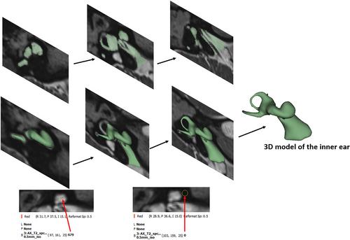
Magnetic resonance imaging (MRI) scans of the temporal bones of candidates for cochlear implants (CIs) enrolled at a tertiary center were retrospectively reviewed from the clinical database. The 3.0-Tesla MRI scans were analyzed using a three-dimensional slicer to visualize the nerve bundles in the internal auditory canal.
Results
A total of 49 ears were analyzed. Twenty ears exhibited normal inner ear anatomy, whereas 29 ears had various inner-ear malformations. The cochlear nerve (CN) was visible on all 20 scans with normal inner-ear anatomy. In addition, the CN was visualized in 18 scans with inner ear malformations. Furthermore, the CN was identified in six of the eight scans with IP type I, whereas in two scans, the CN and vestibular nerve were unclear. Three scans with a common cavity showed only two nerve bundles.
Conclusion
The findings of this study show that the CN can be visualized in most inner-ear anatomical types. Even in severely malformed inner ears, the common nerve bundle that represents the cochlear and vestibular nerves can be visualized. The MRI is highly recommended for CN assessment before CI surgery.

 求助内容:
求助内容: 应助结果提醒方式:


